. 2020 Jan;13(1):27-38.
doi: 10.14802/jmd.19064. Epub 2020 Jan 31.

Principles of Electrophysiological Assessments for Movement Disorders

Affiliations

Principles of Electrophysiological Assessments for Movement Disorders

Kai-Hsiang Stanley Chen et al. J Mov Disord. 2020 Jan.

Abstract

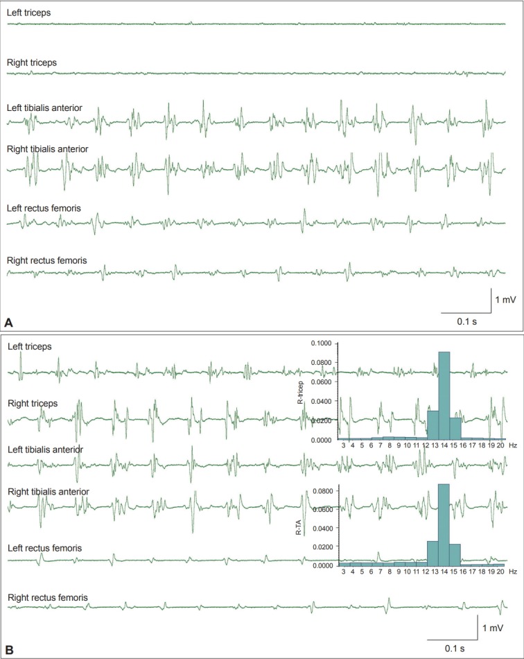
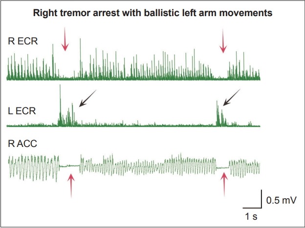
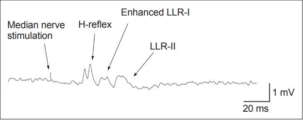
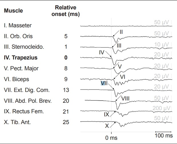
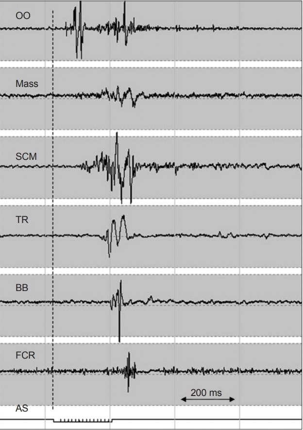
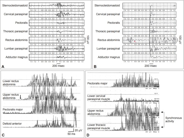
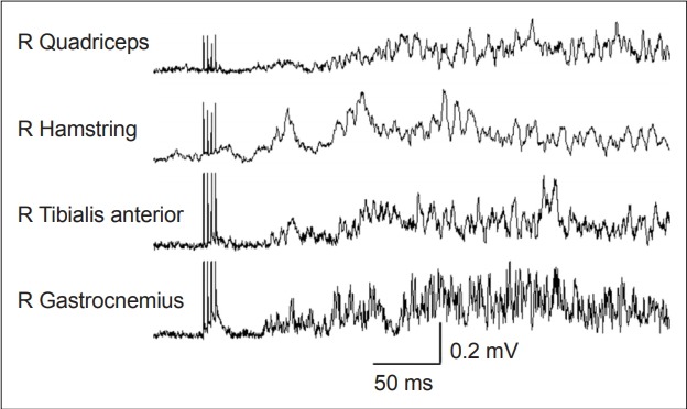
Electrophysiological studies can provide objective and quantifiable assessments of movement disorders. They are useful in the diagnosis of hyperkinetic movement disorders, particularly tremors and myoclonus. The most commonly used measures are surface electromyography (sEMG), electroencephalography (EEG) and accelerometry. Frequency and coherence analyses of sEMG signals may reveal the nature of tremors and the source of the tremors. The effects of voluntary tapping, ballistic movements and weighting of the limbs can help to distinguish between organic and functional tremors. The presence of Bereitschafts-potentials and beta-band desynchronization recorded by EEG before movement onset provide strong evidence for functional movement disorders. EMG burst durations, distributions and muscle recruitment orders may identify and classify myoclonus to cortical, subcortical or spinal origins and help in the diagnosis of functional myoclonus. Organic and functional cervical dystonia can potentially be distinguished by EMG power spectral analysis. Several reflex circuits, such as the long latency reflex, blink reflex and startle reflex, can be elicited with different types of external stimuli and are useful in the assessment of myoclonus, excessive startle and stiff person syndrome. However, limitations of the tests should be recognized, and the results should be interpreted together with clinical observations.

Keywords: Accelerometry; Dystonia; Electroencephalography; Electromyography; Electrophysiology; Myoclonus; Psychomotor disorders.


Conflict of interest statement

Conflict of Interest

The authors have no financial conflicts of interest.

Figures

Figure 1.
Figure 1.
Example of orthostatic tremor. A: Surface electromyography (sEMG) signals recorded from a patient with orthostatic tremor. EMG bursts in the bilateral rectus femoris and tibialis anterior muscles reveal regular firing at a frequency of 14 Hz. B: The patient leaned forward and partially supported his weight with both arms by pressing on a table. Tremors at 14 Hz are now evident in both triceps muscles that are used for support. The inset histograms show the same power spectrum peak at 14 Hz in both the triceps and the tibialis anterior muscles. R-TA: right tibialis anterior muscle.
Figure 2.
Figure 2.
Contralateral ballistic movement induced a transient pause of functional tremor. A case of functional right arm postural tremor. EMG signals were recorded from the bilateral extensor carpi radialis muscles (ECR), and an accelerometer (ACC) was attached to the right middle finger. Transient pauses of the tremors in the right ECR muscle and in the accelerometer recording (red arrows) were observed when the left hand performed voluntary ballistic movements (black arrows).
Figure 3.
Figure 3.
Enhanced long-latency reflex (LLR) in a patient with cortical myoclonus. The long latency reflex showed normal H-reflex and LLR-II responses and enhanced an LLR-I response in a patient with myoclonus. EMG signals were recorded from the abductor pollicis brevis muscle with 20% background activation. Three Hz median nerve stimulations at the motor threshold stimulation intensity were delivered, and a total of 250 trials were recorded.
Figure 4.
Figure 4.
Example of reticular reflex myoclonus due to left medulla compression by the vertebral artery. The myoclonus started in the trapezius muscle with subsequent muscle activation rostrally to the orbicularis oris and caudally to the tibialis anterior muscles. The corresponding onset latency relative to the first contracted trapezius muscle (bold) was shown in ms. The myoclonus has short EMG burst durations. Adapted from Beudel et al.[42] Orb. Oris: orbicularis oris muscle, Sternocleido.: sternocleidomastoid muscle, Pect. Major: pectoralis major muscle, Ext. Dig. Com.: extensor digitorum communis muscle, Abd. Pol. Brev.: Abductor pollicis brevis muscle, Rectus Fem.: rectus femoris muscle, Tib. Ant.: tibialis anterior muscle.
Figure 5.
Figure 5.
Symptomatic hyperekplexia caused by brainstem encephalopathy. EMG recording of the startle response from an unexpected acoustic stimulus (AS, bottom line). The vertical dashed line indicates the beginning of the AS. After the initial blink response, the first muscle response to the AS was from the sternocleidomastoid muscle, with both rostral and caudal spreading. Relatively late EMG responses in the limb muscle and generally long EMG burst durations were the characteristics of the startle reflex. Adapted from van de Warrenburg et al.[47] OO: orbicularis oculi, Mass: master, SCM: sternocleidomastoid, TR: trapezius, BB: biceps brachii, FCR: flexor carpi radialis.
Figure 6.
Figure 6.
Functional propriospinal myoclonus (PSM) and PSM mimicked by healthy subjects. Functional PSM can sometimes be differentiated from idiopathic or symptomatic PSM by (A) the absence of a typical rostral and caudal recruitment order, (B) a burst duration longer than 1,000 ms and isolated muscle activity in the rectus abdominis muscle (red arrow). PSM symptoms can be mimicked. Healthy subjects can mimic (C) typical PSM propagation patterns starting from the upper rectus abdominus muscle (black arrow) and (D) synchronous activation of the truncal flexors and extensors. Adapted from Erro et al.[56] and Kang and Sohn[55].
Figure 7.
Figure 7.
Enhanced exteroceptive reflex in a patient with stiff-person syndrome. The stimulation was delivered to the right tibial nerve, and the EMG signals from the right leg muscles were recorded. Two phases of response were obtained: a brief short latency first phase (about 50 ms), followed by a second phase with a longer latency and duration of more than 100 ms. Adapted from Espay and Chen[70]. R: right.

References

    1. Chen R, Remtulla H, Bolton CF. Electrophysiological study of diaphragmatic myoclonus. J Neurol Neurosurg Psychiatry. 1995;58:480–483. - PMC - PubMed
    1. Mima T, Hallett M, Shibasaki H. Chapter 7. Coherence, cortico-muscular. In: Hallett M, editor. Handbook of Clinical Neurophysiology. Vol 1. Elsevier; 2003. pp. 87–94.
    1. Mauguière F. Chapter 5. Somatosensory evoked responses. In: Hallett M, editor. Handbook of Clinical Neurophysiology. Vol 1. Elsevier; 2003. pp. 45–75.
    1. Pratt H, Politoske D, Starr A. Mechanically and electrically evoked somatosensory potentials in humans: effects of stimulus presentation rate. Electroencephalogr Clin Neurophysiol. 1980;49:240–249. - PubMed
    1. Phielipp NM, Chen R. Chapter 10. Neurophysiologic assessment of movement disorders in humans, in movement disorders. In: LeDoux MS, editor. Movement disorders. 2nd ed. Boston: Academic Press; 2015. pp. 171–186.

LinkOut - more resources