. 2020 Mar;54(2):135-145.
doi: 10.4132/jptm.2019.11.06. Epub 2020 Jan 29.

Colorectal epithelial neoplasm associated with gut-associated lymphoid tissue

Affiliations

Colorectal epithelial neoplasm associated with gut-associated lymphoid tissue

Yo Han Jeon et al. J Pathol Transl Med. 2020 Mar.

Abstract

Background: Colorectal epithelial neoplasm extending into the submucosal gut-associated lymphoid tissue (GALT) can cause difficulties in the differential diagnosis. Regarding GALT-associated epithelial neoplasms, a few studies favor the term "GALT carcinoma" while other studies have mentioned the term "GALT-associated pseudoinvasion/epithelial misplacement (PEM)".

Methods: The clinicopathologic characteristics of 11 cases of colorectal epithelial neoplasm associated with submucosal GALT diagnosed via endoscopic submucosal dissection were studied.

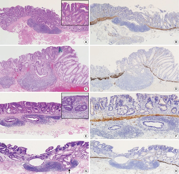
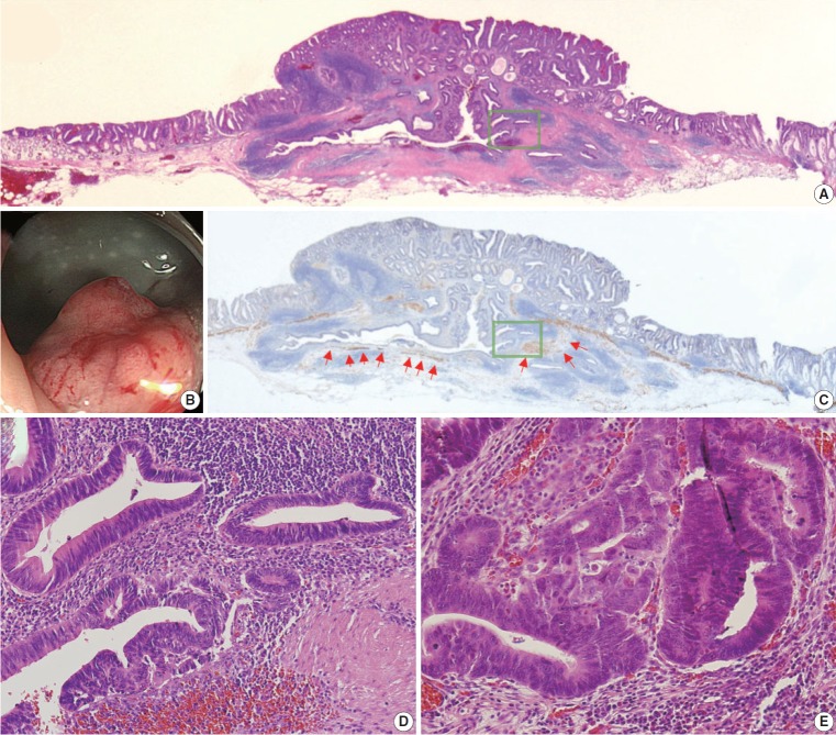
Results: Eight cases (72.7%) were in males. The median age was 59 years, and age ranged from 53 to 73. All cases had a submucosal tumor component more compatible with GALT-associated PEM. Eight cases (72.7%) were located in the right colon. Ten cases (90.9%) had a non-protruding endoscopic appearance. Nine cases (81.8%) showed continuity between the submucosal and surface adenomatous components. Nine cases showed (81.8%) focal defects or discontinuation of the muscularis mucosae adjacent to the submucosal GALT. No case showed hemosiderin deposits in the submucosa or desmoplastic reaction. No case showed single tumor cells or small clusters of tumor cells in the submucosal GALT. Seven cases (63.6%) showed goblet cells in the submucosa. No cases showed oncocytic columnar cells lining submucosal glands.

Conclusions: Our experience suggests that pathologists should be aware of the differential diagnosis of GALT-associated submucosal extension by colorectal adenomatous neoplasm. Further studies are needed to validate classification of GALT-associated epithelial neoplasms.

Keywords: Adenomatous polyps; Colorectal neoplasms; Humans; Lymphoid tissue.


Conflict of interest statement

Conflicts of Interest

The authors declare that they have no potential conflicts of interest.

Figures

Fig. 1.
Fig. 1.
Continuity of submucosal glands with surface adenomatous component and focal defect of muscularis mucosae adjacent to submucosal gut-associated lymphoid tissue (GALT). (A) Case 5. Tubular adenoma with high-grade dysplasia. Histologic continuity of submucosal glands with surface adenomatous component is seen. Submucosal glands with low-grade dysplasia show similar degree of differentiation compared with surface component. However, this neoplasm shows focal area of glands with complex architecture and corresponding high-grade cytologic features in surface mucosa elsewhere (depicted in inset). This case is more compatible with tubular adenoma with high-grade dysplasia than invasive adenocarcinoma, which usually shows less differentiated tumor cells in the deepest part of invasion. (B) Immunohistochemical (IHC) staining for desmin in case 5 shows focal defects of the muscularis mucosae with GALT-associated pseudoinvasion/epithelial misplacement (PEM). (C) Case 9. Tubular adenoma with high-grade dysplasia. Both surface adenomatous component and submucosal glands show high-grade dysplasia and histologic continuity across the muscularis mucosae. (D) IHC staining for desmin in case 9 highlights discontinuous muscularis mucosae. (E) Case 2. Tubular adenoma with high-grade dysplasia. In contrast to cases 5 and 9, no histologic continuity of submucosal glands with surface adenomatous component is seen. Inset depicts glands with high-grade dysplasia in surface mucosa. Narrow rim surrounding submucosal glands is not compatible with typical desmoplasia. Absence of single tumor cells/small clusters of tumor cells, poorly formed or back-to-back glands, solid tumor nests, or “true” desmoplasia favor diagnosis of tubular adenoma with high-grade dysplasia involving GALT (PEM) over adenocarcinoma with “true” submucosal invasion. (F) Intact muscularis mucosae with subjacent GALT of case 2 is identified with IHC staining for desmin. (G) Case 11. Tubular adenoma with low-grade dysplasia. Cystically dilated tumor glands cross through the muscularis mucosae. Note simultaneous crossing over by non-neoplastic glands (indicated by arrow). (H) IHC staining for desmin in case 11. PEM via GALT is accompanied by discontinuous muscularis mucosae rather than hypertrophy of muscularis mucosae.
Fig. 2.
Fig. 2.
Case 3. (A) Histologic continuity along with subtle rimming of muscularis mucosae in submucosa (so-called herniation pattern) favor diagnosis of tubular adenoma with high-grade dysplasia over invasive adenocarcinoma. Depth of neoplasm is more than twice the thickness of surrounding normal colorectal mucosa. (B) Endoscopic appearance of case 3. Exact measurement of neoplasm depth is not available for endoscopist. Superficial neoplastic lesion with height more than one-third of diameter is compatible with the protruding type. (C) Immunohistochemical staining for desmin in case 3. Rimming of muscularis mucosa is indicated by red arrows. (D) Glands with high-grade dysplasia are seen under imaginary line connecting adjacent muscularis mucosa beneath normal mucosa. Panel D corresponds to green boxes of panels A and C. (E) Glands of surface mucosal layer with high-grade dysplasia.

Similar articles

Cited by

References

    1. Neutra MR, Mantis NJ, Kraehenbuhl JP. Collaboration of epithelial cells with organized mucosal lymphoid tissues. Nat Immunol. 2001;2:1004–9. - PubMed
    1. Rubio CA, Puppa G, de Petris G, Kis L, Schmidt PT. The third pathway of colorectal carcinogenesis. J Clin Pathol. 2018;71:7–11. - PubMed
    1. Elmore SA. Enhanced histopathology of mucosa-associated lymphoid tissue. Toxicol Pathol. 2006;34:687–96. - PMC - PubMed
    1. Langman JM, Rowland R. The number and distribution of lymphoid follicles in the human large intestine. J Anat. 1986;149:189–94. - PMC - PubMed
    1. Kealy WF. Colonic lymphoid-glandular complex (microbursa): nature and morphology. J Clin Pathol. 1976;29:241–4. - PMC - PubMed