. 2020 Mar 26;202(8):e00644-19.
doi: 10.1128/JB.00644-19. Print 2020 Mar 26.

HK97 gp74 Possesses an α-Helical Insertion in the ββα Fold That Affects Its Metal Binding, cos Site Digestion, and In Vivo Activities

Affiliations

HK97 gp74 Possesses an α-Helical Insertion in the ββα Fold That Affects Its Metal Binding, cos Site Digestion, and In Vivo Activities

Sasha A Weiditch et al. J Bacteriol. .

Abstract

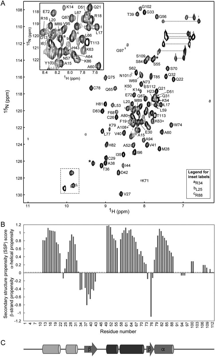
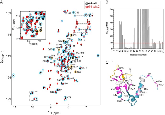
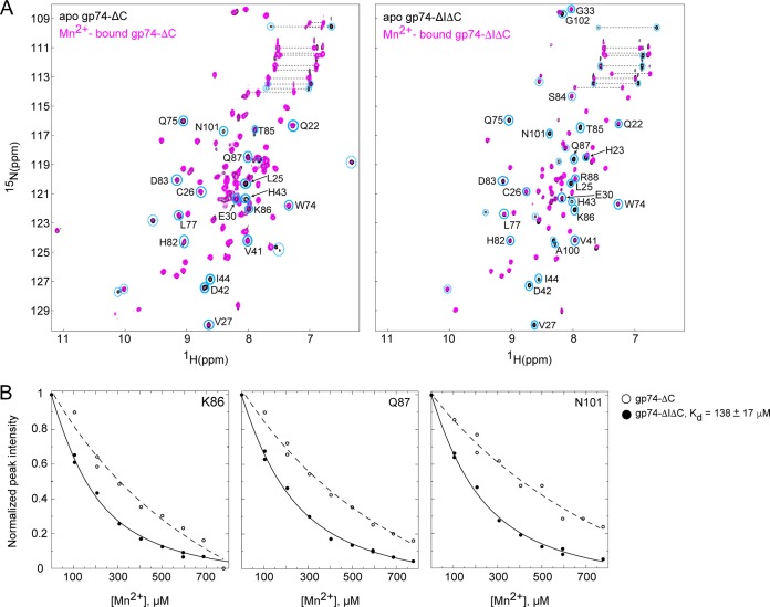
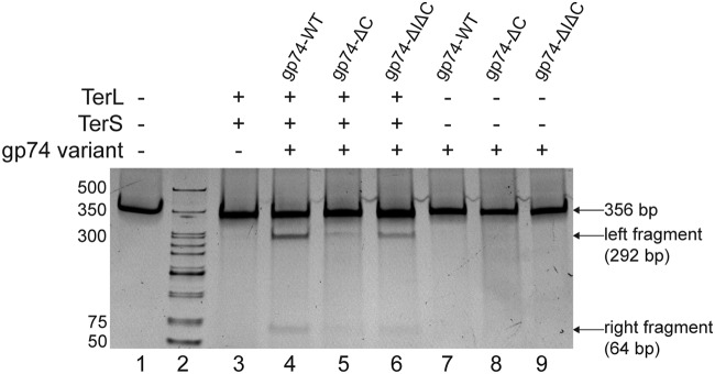
The last gene in the genome of the bacteriophage HK97 encodes gp74, an HNH endonuclease. HNH motifs contain two conserved His residues and an invariant Asn residue, and they adopt a ββα structure. gp74 is essential for phage head morphogenesis, likely because gp74 enhances the specific endonuclease activity of the HK97 terminase complex. Notably, the ability of gp74 to enhance the terminase-mediated cleavage of the phage cos site requires an intact HNH motif in gp74. Mutation of H82, the conserved metal-binding His residue in the HNH motif, to Ala abrogates gp74-mediated stimulation of terminase activity. Here, we present nuclear magnetic resonance (NMR) studies demonstrating that gp74 contains an α-helical insertion in the Ω-loop, which connects the two β-strands of the ββα fold, and a disordered C-terminal tail. NMR data indicate that the Ω-loop insert makes contacts to the ββα fold and influences the ability of gp74 to bind divalent metal ions. Further, the Ω-loop insert and C-terminal tail contribute to gp74-mediated DNA digestion and to gp74 activity in phage morphogenesis. The data presented here enrich our molecular-level understanding of how HNH endonucleases enhance terminase-mediated digestion of the cos site and contribute to the phage replication cycle.IMPORTANCE This study demonstrates that residues outside the canonical ββα fold, namely, the Ω-loop α-helical insert and a disordered C-terminal tail, regulate the activity of the HNH endonuclease gp74. The increased divalent metal ion binding when the Ω-loop insert is removed compared to reduced cos site digestion and phage formation indicates that the Ω-loop insert plays multiple regulatory roles. The data presented here provide insights into the molecular basis of the involvement of HNH proteins in phage DNA packing.

Keywords: HNH endonuclease; NMR spectroscopy; bacteriophage; cos site; divalent metal binding; Ω-loop.


Figures

FIG 1
FIG 1
HK97 gp74 possesses an α-helical insertion within the Ω-loop of the HNH motif. (A) The 15N-1H HSQC spectrum of gp74-WT at 25°C at 600 MHz with a protein concentration of 0.2 mM. Backbone amide cross peaks for which we have resonance assignments have been labeled. For clarity, three resonances in the insert are labeled with the letters a, b, and c to denote the backbone resonances of R35, L25, and R88, respectively. Resonances from the side chain indole 1HN groups of the Trp residues are highlighted with a dashed box. The two resonances of side chain amide groups from each Asn and Gln residue are connected by dotted lines. Side chain Asn and Gln amide groups and Trp indoles have not been assigned or labeled. Resonance assignments for I44 and K50 in gp74-WT were obtained from resonance assignment of gp74-ΔC, as these peaks are of much higher intensity in the gp74-ΔC HSQC spectrum and in spectra from the HCN triple-resonance assignment experiments. The two resonances labeled with asterisks do not possess associated peaks in the triple-resonance data and are thus likely from side chain HN groups. (B) Secondary structure propensity (SSP) values calculated for gp74 averaged over a sliding window of 5 residues. Positive SSP values reflect the α-helical structure for each residue, and negative SSP values reflect the β-strand or extended structure (38). (C) Schematic representation of the secondary structure of gp74, as derived from the SSP values, with α-helices shown as cylinders and β-strands shown as arrows. The two α-helices that form the Ω-loop insert are highlighted in dark gray. The α-helix and β-strands of the ββα-metal fold are shown in the medium gray color, whereas the two N-terminal α-helices are shown in light gray. The β-strands and α-helix of the ββα-metal fold are labeled. Lack of resonance assignments for residues M1 to W12 precludes determining whether these residues form additional secondary structure elements.
FIG 2
FIG 2
Removal of the Ω-loop insert affects residues throughout gp74. (A) Overlay of the two-dimensional 15N-1H HSQC NMR spectra of gp74-ΔC (151 μM) and gp74-ΔIΔC (151 μM). The spectrum of gp74-ΔC is in black and in the background, while the spectrum of gp74-ΔIΔC is in red and in the foreground. As in Fig. 1, Trp side chain indole 1HN resonances are highlighted with a dashed box, and NH2 resonances of Asn and Gln residues are connected by dotted lines. Cyan circles highlight resonances in gp74-ΔC that undergo chemical shift changes with deletion of the Ω-loop insert. Only combined chemical shift changes (Δδtotal) of 9 Hz or more are considered significant (see Materials and Methods). Cyan circles also highlight new resonances that appear in spectra of gp74-ΔIΔC, which are likely derived from residues near the deletion site. Resonances from the Ω-loop insert residues that are missing in the gp74-ΔIΔC spectrum are highlighted by gold circles. Only gp74-ΔC resonances that undergo significant chemical shift changes or that are missing in spectra of gp74-ΔIΔC are labeled in order for comparison with the fully labeled spectrum of gp74-ΔC in Fig. S2 in the supplemental material. (B) The combined chemical shift differences are plotted as a function of residue number. A dashed line depicts the Δδtotal value of 9 Hz. (C) Chemical shift changes associated with removal of the Ω-loop insert are mapped onto the gp74 homology model, which is based on the structure of GVE2 (28), showing only residues in gp74-ΔC (M1 to Y103). The homology model of gp74 is displayed as a ribbon diagram that is colored blue for residues with resonance assignments and gray for residues with no resonance assignments. Pro residues are also colored gray, as these residues do not give signals in NH-based NMR experiments, such as the two-dimensional 1H-15N HSQC experiment. The Ω-loop is colored gold. Because the Ω-loop of GVE2 is smaller than that of gp74 and does not possess α-helices, the Ω-loop in the gp74 homology model is shown as a disordered insert. Further, the position of the insert with respect to the rest of the protein likely also differs from what is observed in the homology model or may change in time on account of the dynamic nature of gp74, as described in the text. However, the similarity in the structures of the two proteins in the ββα fold and N terminal to the ββα fold, as predicted by Phyre2 (49, 50), enables using the gp74 homology model to illustrate the effect of removing the Ω-loop. Cα atoms of residues that exhibit chemical shift changes with removal of the Ω-loop insert are shown as spheres colored from light pink, to highlight the smallest changes, to dark pink for the largest changes. For clarity, only selected residues are labeled.
FIG 3
FIG 3
15N NMR relaxation of gp74-ΔC. 15N R1 and R2 relaxation rates in s−1 are shown as a function of residue number. Secondary structural elements for gp74-ΔC derived from the SSP values are shown at the bottom of the figure.
FIG 4
FIG 4
The Ω-loop insert regulates divalent metal binding to the HNH motif. (A) Overlay of the two-dimensional 15N-1H HSQC NMR spectra of gp74-ΔC (151 μM, left) or gp74-ΔIΔC (151 μM, right) in the absence and presence of Mn2+ (594 μM). The spectra of the apoproteins are in black and in the background, while the spectra of gp74-ΔC or gp74-ΔIΔC with Mn2+ are in magenta and in the foreground. The gp74-ΔC and gp74-ΔΙΔC resonances that undergo significant broadening upon the addition of the paramagnetic Mn2+ ion are highlighted by cyan circles. The solid cyan circles indicate resonances for which we have assignments, while dashed cyan circles highlight resonances that are not assigned. (B) Plots of resonance intensity as a function of Mn2+ concentration for resonances of K86, Q87, and N101 in gp74-ΔC and gp74-ΔIΔC show that gp74-ΔIΔC binds divalent metals with greater affinity than gp74-ΔC. Fitting the data for gp74-ΔIΔC to equation 1 (see Materials and Methods) indicates that gp74-ΔIΔC binds Mn2+ with a Kd value of 138 ± 17 μM. Data for gp74-ΔC could not be used to determine the Kd value for Mn2+ binding on account of the fact that these curves do not reach a plateau at the highest Mn2+ concentrations, indicating a larger Kd value compared to that for the gp74-ΔIΔC/Mn2+ interaction. The curve is shown for gp74-ΔC for comparison with gp74-ΔIΔC.
FIG 5
FIG 5
The Ω-loop insert and C-terminal tail contribute to gp74-mediated enhancement of terminase activity at the HK97 cos site. Digestion assays used 5.0 nM a 356-bp cos DNA, 3.5 μM TerS, 7 μM TerL, and 7 μM gp74 proteins and were visualized on a 4 to 15% acrylamide gel. Digestion of the 356-bp fragment of DNA containing the cos site results in two fragments, the left cohesive fragment (292 bp) and the right cohesive fragment (64 bp). Only a faint band for the left cohesive fragment (292 bp) is observed for the sample containing TerS, TerL, and cos DNA (lane 3). gp74 enhances the terminase-mediated digestion at the cos site, indicated by the visible intensities of both the left and right cohesive fragments (lanes 4, 5, and 6). The intensities of the left and right cohesive fragments are decreased for gp74-ΔC (lane 5) and gp74-ΔIΔC (lane 6). Using the relative intensities of the intact cos DNA band and the left fragment, as the very low intensity of the small right fragment precludes its quantitation, gp74-ΔC and gp74-ΔIΔC decrease cos digestion by 69% ± 15% and 43% ± 17%, respectively, from three separate experiments. Thus, gp74-ΔC and gp74-ΔIΔC are compromised in enhancing the activity of the terminase enzymes toward the cos site.

Similar articles

Cited by

References

    1. Juhala RJ, Ford ME, Duda RL, Youlton A, Hatfull GF, Hendrix RW. 2000. Genomic sequences of bacteriophages HK97 and HK022: pervasive genetic mosaicism in the lambdoid bacteriophages. J Mol Biol 299:27–51. doi:10.1006/jmbi.2000.3729. - DOI - PubMed
    1. Feiss M, Rao VB. 2012. The bacteriophage DNA packaging machine. Adv Exp Med Biol 726:489–509. doi:10.1007/978-1-4614-0980-9_22. - DOI - PubMed
    1. Rao VB, Feiss M. 2015. Mechanisms of DNA packaging by large double-stranded DNA viruses. Annu Rev Virol 2:351–378. doi:10.1146/annurev-virology-100114-055212. - DOI - PMC - PubMed
    1. Kala S, Cumby N, Sadowski PD, Hyder BZ, Kanelis V, Davidson AR, Maxwell KL. 2014. HNH proteins are a widespread component of phage DNA packaging machines. Proc Natl Acad Sci U S A 111:6022–6027. doi:10.1073/pnas.1320952111. - DOI - PMC - PubMed
    1. Moodley S, Maxwell KL, Kanelis V. 2012. The protein gp74 from the bacteriophage HK97 functions as a HNH endonuclease. Protein Sci 21:809–818. doi:10.1002/pro.2064. - DOI - PMC - PubMed

Publication types

Grants and funding

LinkOut - more resources