. 2020 Feb 7;9(2):bio046854.
doi: 10.1242/bio.046854.

Polyphyllin II inhibits liver cancer cell proliferation, migration and invasion through downregulated cofilin activity and the AKT/NF-κB pathway

Affiliations

Polyphyllin II inhibits liver cancer cell proliferation, migration and invasion through downregulated cofilin activity and the AKT/NF-κB pathway

Dejiang Pang et al. Biol Open. .

Erratum in

Abstract

The morbidity and mortality of primary liver cancer is one of the highest amongst all cancers. Deficiency of effective treatment and characteristics of cancer metastasis are believed to be responsible for this situation, thus a great demand is required for new agent development. Polyphyllin II (PP2), an important steroidal saponin extracted from Rhizoma Paris, has emerged as a potential anti-cancer agent, but the effects of PP2 in liver cancers and its underlying mechanisms remain unexplored. In our study, we found that PP2 could remarkably suppress the proliferation of two liver cancer cell lines, HepG2 and BEL7402, resulting in significant cell death. Besides, low doses of PP2 have displayed properties that inhibit cellular motility and invasion of liver cancer cells. In addition, we have found that PP2-mediated cofilin activity suppression was implicated in the inhibition of liver cancer cell motility. Decreased expression of two major hydrolytic enzymes (MMP2/MMP9), through the AKT/NF-κB signaling pathway may also be also responsible for this process. Rescue experiments done with either non-phosphorylatable mutant cofilin-1 (S3A) transfection or an activator of the AKT pathway significantly reversed the inhibition effects of PP2 on liver cancer cells. Taken together, we report a potential agent for liver cancer treatment and reveal its underlying mechanisms.

Keywords: AKT/NF-κB; Cofilin; Liver cancer; MMP2/MMP9; Migration and invasion; Polyphyllin II; Proliferation.


Conflict of interest statement

Competing interestsThe authors declare no competing or financial interests.

Figures

Fig. 1.
Fig. 1.
PP2 suppressed the cell viability and induced cell apoptosis of HepG2 and BEL7402 cells. (A) The structure of Polyphyllin II (PP2). (B,C) Quantifications show the cell viability of HepG2 (B) and BEL7402 (C) cells treated with different concentrations of PP2 for 12 h, 24 h and 36 h. Sorafenib was used as a positive control. (D) Representative images that show the nuclear morphology of HepG2 and BEL7402 cells under PP2 treatment for 24 h labeled with Hoechst 33342. Red arrows show the apoptosis cells. Scale bar: 50μm. (F) Quantification shows that PP2 induces cell death of HepG2 and BEL7402 cells according to Hoechst 33342 staining. ***P<0.001.
Fig. 2.
Fig. 2.
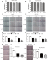
PP2 inhibited cellular motility and invasion of HepG2 and BEL7402 cells. (A,B) Quantifications show the cell viability of HepG2 (A) and BEL7402 (B) cells treated with low doses of PP2. (C–F) Wound healing assay (C,D) and quantifications (E,F) show decreased cellular motility of HepG2 cells and BEL7402 cells treated with 0, 0.5 and 1.0 μM PP2 for 24 and 48 h. (G,I) Cell invasion was analyzed with a Matrigel-coated Boyden chamber. Representative photomicrographs of the membrane-associated cells were assayed by 0.1% Cresyl Violet staining. (H,J) Cell invasion ability was quantitated. **P<0.01, ***P<0.001. Scale bars: 200 μm (C,D); 50 μm (G,I).
Fig. 3.
Fig. 3.
Suppressed cofilin activity was implicated in PP2-inhibited cellular motility of HepG2 and BEL7402 cells. (A–D) Western blots and quantifications show increased phosphorylation of cofilin-1 in the cells treated with 0, 0.5 and 1.0 μM PP2 for 24 h. (E,F) Wound healing assay and quantifications show stably expressed mutant cofilin-1 (S3A) improves cellular motility of HepG2 cells under the treatment of PP2 (1 μM). (G–J) Western blots and quantifications show decreased protein levels of SSH-1 in the cells treated with 0, 0.5 and 1.0 μM PP2 for 24 h. **P<0.01, ***P<0.001. Scale bar: 200 μm.
Fig. 4.
Fig. 4.
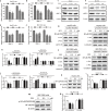
PP2 inhibited the expression of MMP2/MMP9 and the activity of NF-κB in HepG2 and BEL7402 cells. (A,B) The mRNA levels of MMP2/MMP9 were determined with real-time quantitative RT-PCR after the cells were incubated with 0, 0.5 and 1.0 μM PP2 for 24 h. (C–F) Western blots and quantifications show decreased protein levels of MMP2/MMP9 in the cells treated with 0, 0.5 and 1.0 μM PP2 for 24 h. (G–J) Western blots and quantifications show reduced NF-κB protein levels in the nuclear fractions and increased NF-κB protein levels in the cytosolic fractions after treating with 0, 0.5 and 1.0 μM PP2 for 24 h. (K–N) Western blots and quantifications show decreased phosphorylation of NF-κB in the cells treated with 0, 0.5 and 1.0 μM PP2 for 24 h. *P<0.05, **P<0.01, ***P<0.001.
Fig. 5.
Fig. 5.
AKT signaling was implicated in the PP2-suppressed NF-κB/MMPs pathway. (A–D) Western blots and quantifications show reduced pAKT protein levels after treating with 0, 0.5 and 1.0 μM PP2 for 24 h in HepG2 and BEL7402 cells. (E,F) Western blots and quantification show decreased phosphorylation levels of AKT and NF-κB, as well as the expressions of MMP2/MMP9, after PP2 treatment could be rescued by growth factors. (G,H) Wound-healing assay and quantification show cellular motility after PP2 treatment could be rescued by growth factors for 48 h. (I,J) In vitro invasion assays and quantification show invasive ability after PP2 treatment could be rescued by growth factors for 48 h. (K,L) In vitro invasion assays and quantification show invasive ability after PP2 treatment for 48 h could be rescued by activated AKT. (M,N) Western blots and quantification show decreased phosphorylation levels of AKT and NF-κB, as well as the expressions of MMP2/MMP9, after PP2 treatment could be rescued by activated AKT.*P<0.05, **P<0.01, ***P<0.001. Scale bars: 200 μm (G); 50 μm (I,K).

References

    1. Balogh J., Victor D. III, Asham E. H., Burroughs S. G., Boktour M., Saharia A., Li X., Ghobrial R. M. and Monsour H. P. Jr (2016). Hepatocellular carcinoma: a review. J. Hepatocell. Carcinoma 3, 41 10.2147/JHC.S61146 - DOI - PMC - PubMed
    1. Bravo-Cordero J. J., Magalhaes M. A. O., Eddy R. J., Hodgson L. and Condeelis J. (2013). Functions of cofilin in cell locomotion and invasion. Nat. Rev. Mol. Cell Biol. 14, 405-415. 10.1038/nrm3609 - DOI - PMC - PubMed
    1. Charras G. and Paluch E. (2008). Blebs lead the way: how to migrate without lamellipodia. Nat. Rev. Mol. Cell Biol. 9, 730-736. 10.1038/nrm2453 - DOI - PubMed
    1. Chen J.-S., Huang X.-H., Wang Q., Huang J.-Q., Zhang L.-J., Chen X.-L., Lei J. and Cheng Z.-X. (2013). Sonic hedgehog signaling pathway induces cell migration and invasion through focal adhesion kinase/AKT signaling-mediated activation of matrix metalloproteinase (MMP)-2 and MMP-9 in liver cancer. Carcinogenesis 34, 10-19. 10.1093/carcin/bgs274 - DOI - PubMed
    1. Chen Y.-S., He Y., Chen C., Zeng Y., Xue D., Wen F.-Y., Wang L., Zhang H. and Du J.-R. (2014). Growth inhibition by pennogenyl saponins from Rhizoma paridis on hepatoma xenografts in nude mice. Steroids 83, 39-44. 10.1016/j.steroids.2014.01.014 - DOI - PubMed

Publication types

MeSH terms