. 2020 Feb 28;295(9):2687-2697.
doi: 10.1074/jbc.RA119.010540. Epub 2020 Jan 27.

Identification of the growth factor-binding sequence in the extracellular matrix protein MAGP-1

Affiliations

Identification of the growth factor-binding sequence in the extracellular matrix protein MAGP-1

Thomas J Broekelmann et al. J Biol Chem. .

Abstract

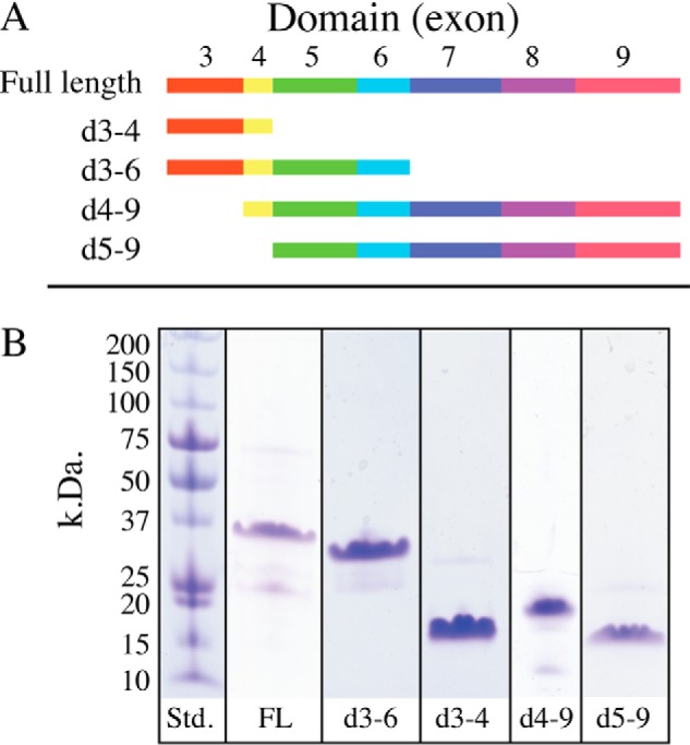
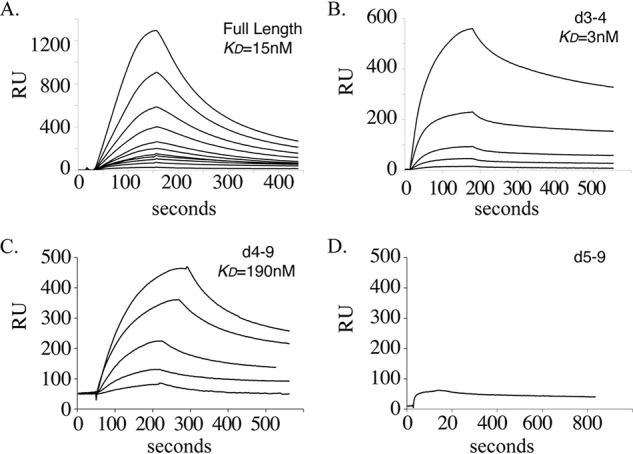
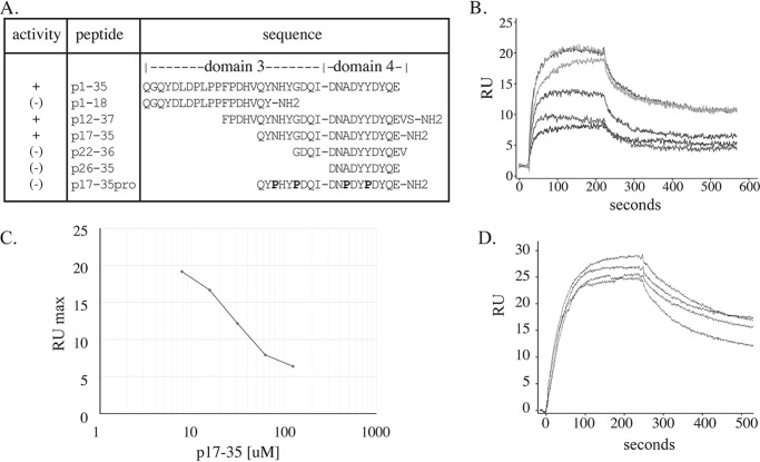
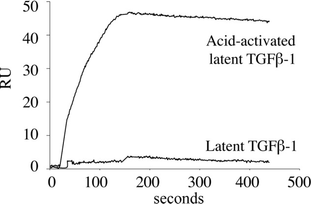
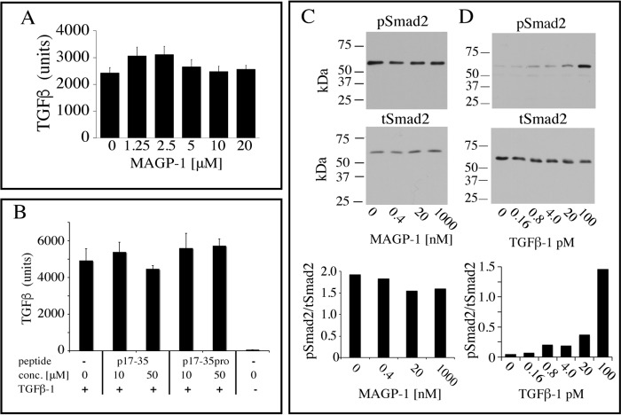
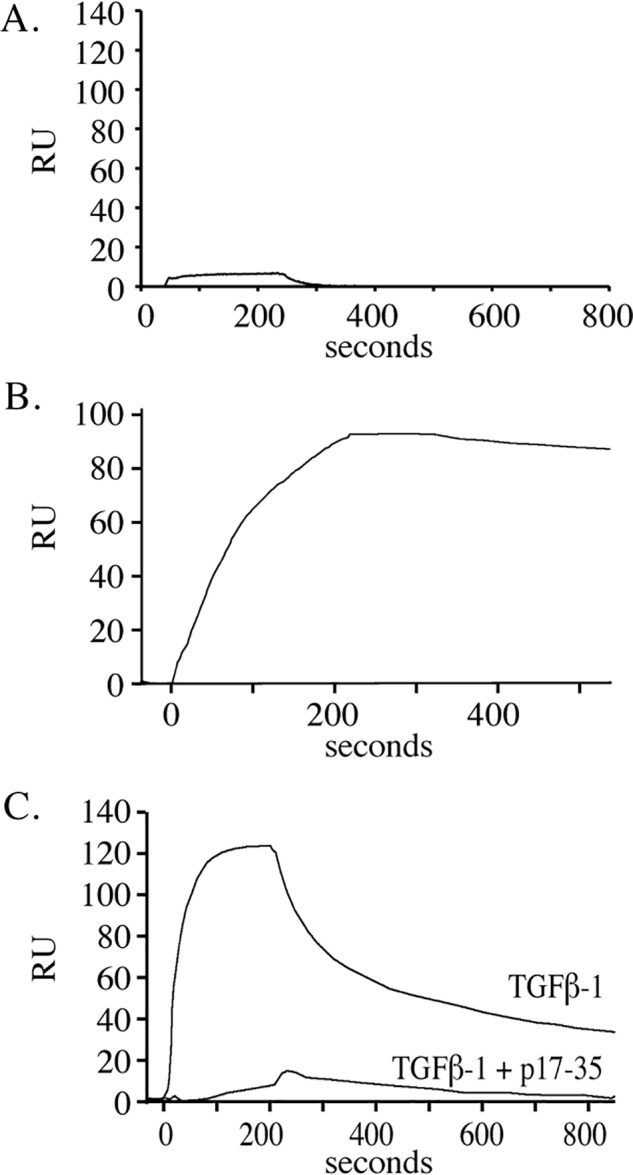
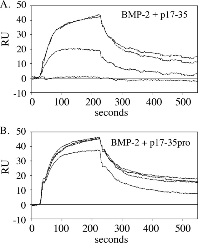
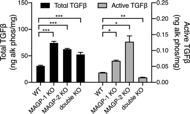
Microfibril-associated glycoprotein-1 (MAGP-1) is a component of vertebrate extracellular matrix (ECM) microfibrils that, together with the fibrillins, contributes to microfibril function. Many of the phenotypes associated with MAGP-1 gene inactivation are consistent with dysregulation of the transforming growth factor β (TGFβ)/bone morphogenetic protein (BMP) signaling system. We have previously shown that full-length MAGP-1 binds active TGFβ-1 and some BMPs. The work presented here further defines the growth factor-binding domain of MAGP-1. Using recombinant domains and synthetic peptides, along with surface plasmon resonance analysis to measure the kinetics of the MAGP-1-TGFβ-1 interaction, we localized the TGFβ- and BMP-binding site in MAGP-1 to a 19-amino acid-long, highly acidic sequence near the N terminus. This domain was specific for binding active, but not latent, TGFβ-1. Growth factor activity experiments revealed that TGFβ-1 retains signaling activity when complexed with MAGP-1. Furthermore, when bound to fibrillin, MAGP-1 retained the ability to interact with TGFβ-1, and active TGFβ-1 did not bind fibrillin in the absence of MAGP-1. The absence of MAGP was sufficient to raise the amount of total TGFβ stored in the ECM of cultured cells, suggesting that the MAGPs compete with the TGFβ large latent complex for binding to microfibrils. Together, these results indicate that MAGP-1 plays an active role in TGFβ signaling in the ECM.

Keywords: MFAP2; TGFβ; bone morphogenetic protein (BMP); cell signaling; extracellular cellular matrix (ECM); extracellular matrix; fibrillar; fibrillin; growth factor; growth factor signaling; microfibril; microfibril associated glycoprotein (MAGP); protein-protein interaction; transforming growth factor beta (TGFβ).


Conflict of interest statement

The authors declare that they have no conflicts of interest with the contents of this article

Figures

Figure 1.
Figure 1.
Domain structure of MAGP-1 and purity of expressed peptides. A, full-length, secreted, MAGP-1 protein is encoded by seven exons (exons 3–9). In this report, each exon defines a protein domain, and truncations used for functional analysis were constructed based on exon boundaries. The schematic shows the domains included in the recombinant fragments (d followed by exon numbers). B, SDS-PAGE analysis of recombinant full-length MAGP-1 (FL) and purified truncated peptides shown in panel A. Proteins were separated on 8–25% Phastgels under reducing conditions and detected with Coomassie Brilliant Blue. The observed and expected molecular weights of each fragment are described in “Results.”
Figure 2.
Figure 2.
Binding of TGFβ-1 to purified full-length MAGP-1 or MAGP-1 fragments. A, graph of isotherms from the binding of TGFβ-1 to a CM-5 chip coated with full-length MAGP-1 (2200 resonance units coupled). TGFβ-1 concentrations were, from the top, 100, 66.7, 44.4, 29.6, 19.8, 13.2, 8.8, 5.9, 3.9, 2.6, and 1.7 nm. B–D, interaction of TGFβ-1 (400, 200, 80, 67, and 27 nm) with 440 resonance units of MAGP-1 domain protein 3–4 (B), with 4300 resonance units of MAGP-1 domain 4–9 (TGFβ-1 concentrations of 120, 60, 48, 24,12 nm) (C), and 400 nm TGFβ-1 with 2140 resonance units of MAGP-1 domain 5–9 (D).
Figure 3.
Figure 3.
Fine mapping of TGFβ-1 binding activity in domains 3–4. A, summary of the inhibitory activity, the sequence, and the location of the peptides tested for inhibition of TGFβ-1 (50 nm) binding to 120 resonance units (RU) of full-length MAGP-1 coupled to a carboxymethyl dextran hydrogel surface sensor chip (MAGP-1 chip). B, a graph of isotherms showing p17–35 dose-dependent inhibition of TGFβ-1 binding to the MAGP-1–coated chip. C, relates the maximal binding of TGFβ-1 (at 100 s) to the MAGP-1 chip in the presence of increasing concentrations of peptide p17–35 (taken from panel B). D, isotherms show that the control peptide p17–35pro (100, 50, 25, and 0 μm) has no inhibitory effect on TGFβ-1 binding to MAGP-1.
Figure 4.
Figure 4.
MAGP-1 does not bind latent TGFβ-1. Latent TGFβ-1 (15 nm) was injected onto a MAGP-1 chip, and no interaction was detected. A second sample of latent TGFβ-1 was treated with 100 mm HCl for 10 min at room temperature, neutralized with 100 mm NaOH, and then diluted to a final concentration of 15 nm. The binding isotherm shows that the acid-activated TGFβ-1 bound with high affinity to the MAGP-1 chip. The injection volume was 125 μl, and the flow rate was 50 μl/min for both injections.
Figure 5.
Figure 5.
MAGP-1 and p17–35 do not block TGFβ-1 signaling. A, active TGFβ-1 (80 pm) was mixed with the indicated concentrations of MAGP-1, incubated for 15 min, and then added to MFB-F11 cells for 24 h. The culture medium was then assayed for alkaline phosphatase activity, which provides a measure of TGFβ activity. Shown are mean ± S.D., n = 8. B, active TGFβ-1 (80 pm) was mixed with 10 μm or 100 μm p17–35, p17–35pro, or no peptide for 15 min and added to MFB-F11 cells for 24 h. A control with no added TGFβ-1 was included. The medium was collected and assayed for alkaline phosphatase activity. Shown are mean ± S.D., n = 4. One-way analysis of variance showed no differences between samples in panels A or B. C, increasing concentrations of soluble MAGP-1 were pre-incubated for 1 h with 50 pm TGFβ-1 and the complex incubated with RFL-6 cell (a fibroblast line that does not make MAGP-1) for 15 min. Cell extracts were then analyzed by Western blotting for phospho-Smad2 (pSmad-2) (top) and for total Smad2 (tSmad2) (bottom). The ratio of pSmad2 to tSmad2 is shown in the bottom graph. D, Smad2 phosphorylation in RFL-6 cells is TGFB-1 dose dependent with minimal baseline phosphorylation. RFL-6 cells were treated with the indicated dose of TGFβ-1 and levels of phosphorylated Smad-2 (top panel) or total Smad-2 (bottom panel) were determined from cell extracts by Western blot analysis. The ratio of pSmad2 to tSmad2 is shown in the bottom graph.
Figure 6.
Figure 6.
The structure of TGFβ-1 with and without the MAGP-1 p17–35 peptide. Top, side and top views of the TGFβ-1 dimer with solvent exposed residues Arg-94, Lys-95, and Lys-97 shown in green. Bottom, representative binding of the MAGP-1 p17–35 peptide (shown in black) to TGFβ-1. The MAGP-1 peptide remains largely disordered while consistently interacting with the positive residues in the TGFβ-1 finger region.
Figure 7.
Figure 7.
TGFβ-1 binds to fibrillin-2 only in the presence of MAGP-1. A, purified full-length fibrillin-2 was covalently coupled (3000 resonance units) to a carboxymethyl dextran hydrogel surface sensor chip (fibrillin-2 chip), and TGFβ-1 was injected at a concentration of 50 nm at 25 μl per minute for 3.5 min. Data show no measurable interaction between the two proteins. B, MAGP-1 (6.2 nm for 3.5 min at 25 μl/min) bound to fibrillin-2 with an apparent affinity of 100 pm. C, TGFβ-1 (50 nm for 3.5 min at 25 μl/min) bound to the MAGP-1-fibrillin-2 complex whereas TGFβ-1 pre-incubated with 50 μm p17–35 peptide failed to bind to the complex.
Figure 8.
Figure 8.
BMP-2 binding localizes to the same domain on MAGP-1 as TGFβ-1. A, isotherms documenting binding of 50 nm BMP-2 to a MAGP-1-coated chip. B, binding was inhibited by preincubation of BMP-2 with peptide p17–35 from the top 0, 5, 25, and 50 uM) but not the control peptide p17–35pro (0, 5, 25, and 50 uM).
Figure 9.
Figure 9.
MAGP 1 and MAGP-2 modulate TGFB levels in fibroblast ECM. Comparison of total TGFβ levels (black bars) in 14-day post confluent WT and MAGP knockout cell layers showing significant increases in total TGFβ in MAGP-1 knockout (**, p > 0.01, n = 3), MAGP-2 knockout (*, p > 0.05, n = 3), and MAGP-1:MAGP-2 double knockout (**, p > 0.01) fibroblasts. Similarly, active TGFβ levels (gray bars) are significantly increased in MAGP-1 knockout (*, p > 0.05, n = 2) and MAGP-2 knockout (*, p > 0.05, n = 3) cells, but are decreased (**, p > 0.01 n = 3) in the double knockouts.

References

    1. Hynes R. (2009) The extracellular matrix: Not just pretty fibrils. Science 326, 1216–1219 10.1126/science.1176009 - DOI - PMC - PubMed
    1. Robertson I., Jensen S., and Handford P. (2011) TB domain proteins: Evolutionary insights into the multifaceted roles of fibrillins and LTBPs. Biochem. J. 433, 263–276 10.1042/BJ20101320 - DOI - PubMed
    1. Davis E. C., Roth R. A., Heuser J. E., and Mecham R. P. (2002) Ultrastructural properties of ciliary zonule microfibrils. J. Struct. Biol. 139, 65–75 10.1016/S1047-8477(02)00559-2 - DOI - PubMed
    1. Goldfischer S., Coltoff-Schiller B., and Goldfischer M. (1985) Microfibrils, elastic anchoring components of the extracellular matrix, are associated with fibronectin in the zonule of Zinn and aorta. Tissue Cell 17, 441–450 10.1016/0040-8166(85)90023-0 - DOI - PubMed
    1. Wagenseil J. E., and Mecham R. P. (2009) Vascular extracellular matrix and arterial mechanics. Physiol. Rev. 89, 957–989 10.1152/physrev.00041.2008 - DOI - PMC - PubMed

Publication types

LinkOut - more resources