Clinical Trial
. 2020 Feb;26(2):200-206.
doi: 10.1038/s41591-019-0735-5. Epub 2020 Jan 27.

Lentiviral gene therapy for X-linked chronic granulomatous disease

Collaborators, Affiliations
Clinical Trial

Lentiviral gene therapy for X-linked chronic granulomatous disease

Donald B Kohn et al. Nat Med. 2020 Feb.

Abstract

Chronic granulomatous disease (CGD) is a rare inherited disorder of phagocytic cells1,2. We report the initial results of nine severely affected X-linked CGD (X-CGD) patients who received ex vivo autologous CD34+ hematopoietic stem and progenitor cell-based lentiviral gene therapy following myeloablative conditioning in first-in-human studies (trial registry nos. NCT02234934 and NCT01855685). The primary objectives were to assess the safety and evaluate the efficacy and stability of biochemical and functional reconstitution in the progeny of engrafted cells at 12 months. The secondary objectives included the evaluation of augmented immunity against bacterial and fungal infection, as well as assessment of hematopoietic stem cell transduction and engraftment. Two enrolled patients died within 3 months of treatment from pre-existing comorbidities. At 12 months, six of the seven surviving patients demonstrated stable vector copy numbers (0.4-1.8 copies per neutrophil) and the persistence of 16-46% oxidase-positive neutrophils. There was no molecular evidence of either clonal dysregulation or transgene silencing. Surviving patients have had no new CGD-related infections, and six have been able to discontinue CGD-related antibiotic prophylaxis. The primary objective was met in six of the nine patients at 12 months follow-up, suggesting that autologous gene therapy is a promising approach for CGD patients.


Conflict of interest statement

Competing interests

D.B.K. (Kohn), H.L.M., D.A.W. & A.J.T. are Scientific Advisory Board members and H.B.G. is Chief Scientific Officer for Orchard Therapeutics. H.B.G. is an employee and equity/stock holder for Orchard Therapeutics. A.J.T. is an equity/stock holder for Orchard Therapeutics. Orchard Therapeutics has obtained an exclusive option to license from Genethon for the rights and know-how related to the lentiviral vector G1XCGD. C.A.B. and T.P. consult for a sequencing service provider, and C.A.B. consults for Novimmune and SOBI. Eurofins Genomics Sequencing Europe (formerly GATC Biotech AG) is a for-profit company (sequencing service provider). The work performed by Eurofins Genomics Sequencing Europe included in the manuscript is provided to the greater scientific community as a fee for service product. E.C.M. reports Advisory Board attendance for Orchard Therapeutics.

With regards to interests outside of the submitted work, L.D.W. reports grants from the St. Baldrick's Foundation, Damon-Runyon Cancer Research Foundation, and Alex's Lemonade Stand Foundation and personal fees from Magenta Therapeutics. A.J.T. reports Board membership and consultancy with Rocket Pharmaceuticals, Generation Bio, and Board membership with 4BIOCapital. S.-Y.P. reports salary support from Boston Children’s Hospital and a grant from National Institutes of Health. K.L.S. reports personal fees and non-financial support from Orchard Therapeutics, Ltd. E.M.K., G.S., M.A., K.F.B., U.C., S.S.D.R., M.J.D., C.Y.K., D.L.-R., C.R., N.I., K.G., K.S., J.D., E.C.M., D.B.K. (Kuhns), J.G., H.R., J.K.E., G.H., P.E.N., F.D.B., M.G., J.X.-B.D. and A.G. have no competing interests to disclose.

Figures

Extended Data Fig. 1
Extended Data Fig. 1. Schematic representation of the transfer plasmid pCCL_chimGP91_WPRE_Kana
CMV promoter; R, R region of long terminal repeat (LTR); U5, Region unique to the 5’ LTR; Psi, encapsidation sequence; RRE, rev responsive element; cPPT, central polypurine tract; CTSG, cathepsin G 5’ minimal flanking regions; FES, human c-fes; coGP91, codon-optimized sequence CYBB gene which is translated into the cytochrome b-245, beta polypeptide; WPRE, mutated woodchuck hepatitis virus posttranscriptional regulatory element; PPT, HIV-1 polyadenylated region (post translational), dU3, region unique to the 3’ LTR (U3) of the HIV-1 LTR, in which a 400 bp deletion (d) was introduced to inactivate the HIV-1 enhancer/promoter region; SV40 ori, Simian Virus 40 origin of replication; KANr, kanamycin resistance gene; COLE1, Colicin E1 resistance gene.
Extended Data Fig. 2
Extended Data Fig. 2. Cell products and busulfan conditioning
Panel a shows the final CD34+ cell products (left-axis) obtained for each patient. Panel b shows the VCN per infused cell post-transduction as determined by quantitative PCR. Transduction was also assessed by analyzing the percentage of vector CFUs PCR-positive for GX1CGD vector detected in the final cell product (Panel c). Vertical bars indicate mean and SD. Panel d displays the total net AUC busulfan exposure obtained by the sum of all pharmacokinetic measurements. Target total exposure of 70,000–75,000 ng/mL*h is indicated by the shaded area. AUC, area under the curve; CFU, colony-forming unit; PCR, polymerase chain reaction; SD, standard deviation; VCN, vector copy number
Extended Data Fig. 3
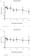
Extended Data Fig. 3. Neutrophil DHR and VCN 24 months post treatment
Panel a shows the mean (±SEM) of % DHR neutrophil up to 24 months post treatment, as measured by a dihydrorhodamine oxidation assay. Panel b shows the mean neutrophil VCN (±SEM) up to 24 months post treatment. DHR, dihydrorhodamine; SEM, standard error of mean; VCN, vector copy number
Extended Data Fig. 4
Extended Data Fig. 4. X-CGD lineage VCN
VCN determined by qPCR in multiple-cell lineages for patients surviving >1 month post-GT at each follow up, showing stable integration of vector into multiple immune cell types. GT, gene therapy; qPCR, quantitative PCR; VCN, vector copy number
Extended Data Fig. 5
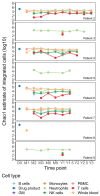
Extended Data Fig. 5. Longitudinal analysis of inferred minimal population sizes
Longitudinal analysis of minimal population size estimated using the CHAO1 method. The x-axis shows time since cell infusion. D0 indicates the pre-infusion product. The y-axis shows the numbers of unique integration sites (log scale). Cell types are color coded (bottom). For a few patients a reduced number of cell types were available for analysis. NK, natural killer; PBMC, peripheral blood mononuclear cell
Extended Data Fig. 6
Extended Data Fig. 6. Catalogue of vector integration sites at genes of concern
Catalogue of cell clones with integration sites within MECOM (MDS/EVI1), PRDM16, and SETBP2. These genes were chosen for analysis because they were targets of integration in expanded cell clones in the first-RV-based CGD trial, and were implicated in adverse outcomes. The x-axis shows the time point queried, the y-axis shows clonal abundance. Cell types queried are color coded (key at the bottom). No cell clones with integration sites near these genes of concern reaches ≥0.3% in abundance and there is no evidence for longitudinal increases in proportion. CGD, chronic granulomatous disease; γ-RV, gammaretroviral; NK, natural killer; PBMC, peripheral blood mononuclear cell
Fig. 1
Fig. 1. Materials and methods (vector map and procedures)
Panel a displays the schematic representation of the G1XCGD lentiviral vector (LV) used to transduce CD34+ peripheral blood and bone marrow stem and progenitor cells in which expression of a codon-optimized human CYBB cDNA encoding for gp91phox is controlled by a chimeric regulatory element containing the Cathepsin G and Cfes gene promoter/enhancers, with a downstream WPRE to boost expression. Panels b and c show the schemas for the fresh cell and frozen/cryopreserved cell procedures, respectively. Differences between the two procedures include the timing of the primary harvest and back-up ‘rescue’ harvest of CD34+ cells, the timing of G1XCGD vector addition, and that the cryopreservation procedure allowed full cell product characterization and release criteria to be met before cytoreductive conditioning was performed. AUC, area under the curve; BM, bone marrow; CatG/Cfes, CTSG encoding Cathepsin G and the FES gene encoding Cfes; COA, certificate of analysis; G-CSF, granulocyte colony-stimulating factor; LV, lentiviral vector; mPB, mobilized peripheral blood; WPRE, Woodchuck hepatitis virus post-transcriptional regulatory element
Fig. 2
Fig. 2. Biochemical and clinical evidence of successful engraftment
Panel a shows the nitro-blue tetrazolium (NBT) test to detect functional circulating neutrophils from a peripheral blood sample from Patient 2 pre-gene therapy (left) and at 3 months after receiving gene therapy with G1XCGD (right). Generation of superoxide leads to reduction of NBT and formation of dark blue formazan precipitates in the observed cells. The scale bar measures approximately 10 micrometres; mature human neutrophils on a blood smear have an average diameter of 12–15 micrometres. Panel b shows the results of DHR fluorescence flow cytometry assaying functional oxidase activity in neutrophils over a 2-year period in Patient 3 post-gene therapy, and in a healthy control. After neutrophil stimulation with PMA (right), the fraction of DHR+ neutrophils were quantified. The percentage of DHR+ neutrophils in all treated patients at each time point is shown in Panel c. NAPDH-oxidase activity was quantified by measurement of neutrophil-stimulated reduction of ferricytochrome c, corrected for the percentage of oxidase-positive cells. Data for Patients 2, 4 and 7 are shown in Panel d following 10 minutes and 60 minutes of stimulation with PMA. The lower limit of normal superoxide generation for each timepoint is indicated by the dashed line. Panel e shows the neutrophil VCN for all patients measured at each time point for which data were available. VCN remained stable for six of seven surviving patients but decreased over time for one patient (Patient 5), who remains clinically well at follow-up with antimicrobial support. Panel f shows the percentage of DHR+ neutrophils versus granulocyte VCN for the seven surviving patients at 12 months. R2=0.44; the dashed line represents 10% DHR. NBT, nitroblue tetrazolium; DHR, dihydrorhodamine; GT, gene therapy; NL, normal; PMA, phorbol myristate acetate; VCN, vector copy number
Fig. 3
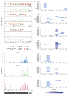
Fig. 3. Analysis of vector integration-site distributions and promotor methylation
Panel a shows longitudinal analysis of unique cell clones contributing to each cell type, inferred from counts of unique integration sites. The x-axis shows time since cell infusion. D0 indicates the pre-infusion product. The y-axis shows the numbers of unique integration sites (log scale). Cell types are color coded (bottom). For a few patients a reduced number of cell types were available for analysis. Panel b is a heat map illustrating the most abundant clones in each patient and their longitudinal behavior. Neutrophils were selected for this analysis because previous adverse events in CGD gene therapy with γ-RV vectors involved outgrowth of myeloid cells. The x-axis shows the time post-treatment. The rows show cell clones, named by the nearest human gene (labels on left of figure). The relative abundance is shown by the heat map scale (bottom of figure). Quantification was carried out using fragment lengths to estimate abundance . In Panel c, methylation of CpG dinucleotides is shown in Patients 2, 3 and 9 at 2.5 years, 18 months and 9 months post-gene therapy, respectively. The x-axis shows the positions of CpG dinucleotides relative to the gp91 mini-gene. The y-axis shows the percentage of methylation at each position. The methylation levels across the CpG islands are low for all samples, indicating that the gp91 mini-gene is not transcriptionally repressed. CGD, chronic granulomatous disease; MSP, myeloid-specific promoter; NK, natural killer; PBMC, peripheral blood mononuclear cell; PCR, polymerase chain reaction; VISA, longitudinal vector integration-site analysis.

References

    1. Winkelstein JA, et al. Chronic granulomatous disease. Report on a national registry of 368 patients. Medicine (Baltimore) 2000;79:155–169. - PubMed
    1. Holland SM. Chronic granulomatous disease. Hematol Oncol Clin North Am. 2013;27:89–99. doi: 10.1016/j.hoc.2012.11.002. viii. - DOI - PMC - PubMed
    1. Gungor T, et al. Reduced-intensity conditioning and HLA-matched haemopoietic stem-cell transplantation in patients with chronic granulomatous disease: a prospective multicentre study. Lancet. 2014;383:436–448. doi: 10.1016/S0140-6736(13)62069-3. - DOI - PubMed
    1. Parta M, et al. Allogeneic reduced-iIntensity hematopoietic stem cell transplantation for chronic granulomatous disease: a single-center prospective trial. J Clin Immunol. 2017;37:548–558. doi: 10.1007/s10875-017-0422-6. - DOI - PMC - PubMed
    1. Ott MG, et al. Correction of X-linked chronic granulomatous disease by gene therapy, augmented by insertional activation of MDS1-EVI1, PRDM16 or SETBP1. Nat Med. 2006;12:401–409. doi: 10.1038/nm1393. - DOI - PubMed

Publication types

MeSH terms