Case Reports
. 2019 Dec 19;11(12):e6418.
doi: 10.7759/cureus.6418.

A Multidisciplinary Approach in the Management of a Paediatric Posterior Fossa Ischaemic Stroke: A Case Report

Affiliations
Case Reports

A Multidisciplinary Approach in the Management of a Paediatric Posterior Fossa Ischaemic Stroke: A Case Report

Orazio Buonomo et al. Cureus. .

Abstract

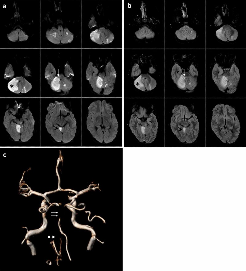
Posterior circulation acute ischaemic stroke in childhood is a rare but life-threatening disease. We describe a paediatric case of a 17-year-old Indian boy who was admitted to our centre for headache, nausea, vomiting, asthenia, and fever for two days. Computed tomography angiography (CTA), magnetic resonance angiography (MRA) and digital subtraction angiography (DSA) were performed, showing a thrombotic occlusion of the basilar artery due to focal dissection into the proximal third of the left vertebral artery. In a multidisciplinary fashion, we decided to perform a direct aspiration first pass technique (ADAPT), which led to the complete recanalization of either the left vertebral artery or the basilar artery. Twenty-four hours later, despite the anti-edemigenic medical therapy, a preventive occipital craniectomy was performed because of the presence of cerebral oedema to avoid the possible worsening of the patient and compression on the brainstem. Our experience emphasizes the importance of a multidisciplinary and preventive approach in the management of a paediatric posterior fossa ischaemic stroke.

Keywords: basilar artery occlusion; endovascular treatment; multidisciplinary approach in stroke care; occipital craniectomy; paediatric ischaemic stroke; posterior fossa ischaemic stroke; vertebral artery dissection.


Conflict of interest statement

The authors have declared that no competing interests exist.

Figures

Figure 1
Figure 1. Admission magnetic resonance imaging (MRI)
An MRI scan taken upon the patient’s arrival that demonstrates: a) a hyperintense hyperacute ischaemic area in the right cerebellar hemisphere (black star) and in the right-sided vermis (black arrowhead) with partial involvement of the right middle cerebellar peduncle and the upper cerebellar peduncle (white arrow) in diffusion-weighted imaging (DWI) sequence; b) a hyperintense acute ischaemic area in the right cerebellar hemisphere (black star) and in the right-sided vermis (black arrowhead) with partial involvement of the right middle cerebellar peduncle and the upper cerebellar peduncle (white arrow) in fluid-attenuated inversion recovery (FLAIR) sequence; c) magnetic resonance angiography (MRA) shows the absence of flow in the middle-distal tract of the basilar artery (double white arrows) with a reduction similar to that of both vertebral arteries (white arrow with dot)
Figure 2
Figure 2. Endovascular treatment
The digital substraction angiogram (DSA) images demonstrate the various phases of treatment: a) an anteroposterior (AP) angiogram of the right vertebral artery shows the common origin (black arrow) of the artery with a costocervical trunk; b) AP angiograms of the left vertebral artery in the neck and intracranial areas show a focal length dissection of the proximal V2 trait (double black arrows) and (c) thrombotic occlusion (black arrow with dot) immediately adjacent to the origin of the posterior inferior cerebellar artery (PICA); d-e) AP and latero-lateral (LL) angiograms of the left vertebral artery after the first pass with the catalyst aspiration catheter show the basilar artery occluded at the apex (black arrowheads); f-g) AP and LL angiograms of the left vertebral artery after the second pass with the catalyst aspiration catheter demonstrate normal posterior circulation (modified thrombolysis in cerebral infarction (mTICI 3)); h) AP angiogram of the left vertebral artery in the neck shows the stability of the focal length dissection of the proximal V2 trait (black star) with a regular and orthograde flow.
Figure 3
Figure 3. Computed tomography (CT) and magnetic resonance imaging (MRI) examination after endovascular treatment
a) Six hours after treatment, a CT scan showed the previously diagnosed ischaemic outbreaks in addition to initial dilatation of the supratentorial ventricular system (black arrowhead) and a reduction of the Silvio aqueduct and the fourth ventricle (black star), due to cerebral cytotoxic oedema; b) 24 hours after the endovascular procedure, magnetic resonance angiography (MRA) showed typical posterior circulation; c) in diffusion-weighted imaging (DWI) sequence and d) in fluid-attenuated inversion recovery (FLAIR) sequence, the ischaemic lesion (black stars) is evident with the same findings as those seen in the last CT scan. Immediately after MRI, a preventive occipital craniectomy was performed.
Figure 4
Figure 4. Computed tomography (CT) scan five days after neurosurgical treatment
CT scan five days after occipital craniectomy (black stars) demonstrates physiological visualization of the cranial subarachnoid spaces due to the reduction of the compressive effect (black arrows).

References

    1. Pediatric arterial ischemic stroke: epidemiology, risk factors, and management. Felling RJ, Sun LR, Maxwell EC, Goldenberg N, Bernard T. Blood Cells Mol Dis. 2017;67:23–33. - PubMed
    1. Global, regional, and country-specific lifetime risks of stroke, 1990 and 2016. GBD 2016 Lifetime Risk of Stroke Collaborators, Feigin VL, Nguyen G, et al. N Engl J Med. 2018;379:2429–2437. - PMC - PubMed
    1. Endovascular mechanical thrombectomy for acute stroke in young children. Sun LR, Felling RJ, Pearl MS. J Neurointerv Surg. 2019;11:554–558. - PubMed
    1. Extracranial vertebral artery dissection in children: natural history and management. Simonnet H, Deiva K, Bellesme C, et al. Neuroradiology. 2015;57:729–738. - PubMed
    1. Childhood posterior circulation arterial ischemic stroke. Mackay MT, Prabhu SP, Coleman L. Stroke. 2010;41:2201–2209. - PubMed

Publication types

LinkOut - more resources