

Implantation of allogenic umbilical cord blood-derived mesenchymal stem cells improves knee osteoarthritis outcomes: Two-year follow-up
- PMID: 31988992
- PMCID: PMC6965506
- DOI: 10.1016/j.reth.2019.10.003
Implantation of allogenic umbilical cord blood-derived mesenchymal stem cells improves knee osteoarthritis outcomes: Two-year follow-up
Abstract
Introduction: Clinical outcomes after the implantation of allogenic human umbilical cord blood-derived mesenchymal stem cells (hUCB-MSCs) in osteoarthritic knees have been rarely reported. Our study aimed to investigate clinical outcomes of osteoarthritic patients who underwent hUCB-MSC implantation.
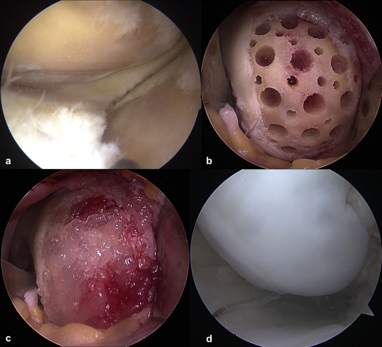
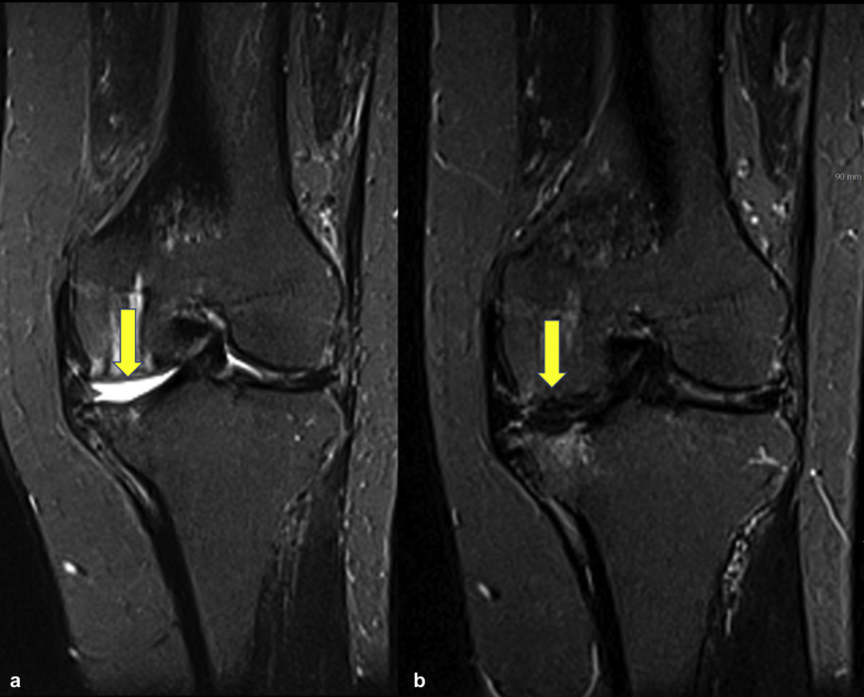
Methods: In this case series (level of evidence: 4), from January 2014 to December 2015, 128 patients with full-thickness cartilage lesions (International Cartilage Repair Society grade 4 and Kellgren-Lawrence grade ≤3) who underwent hUCB-MSC implantation were retrospectively evaluated with a minimum of 2-year follow-up. After removing the sclerotic subchondral bone with an arthroscopic burr, 4-mm-diameter holes were created at 2-mm intervals, and hyaluronic acid and hUCB-MSCs were subsequently mixed and implanted in the holes and other articular defect sites.Clinical outcomes were evaluated preoperatively, 1 year postoperatively, and 2 years postoperatively (minimum) using visual analog scale (VAS), Western Ontario and McMaster Universities Osteoarthritis Index (WOMAC), and International Knee Documentation Committee (IKDC) scores. To assess clinical outcomes, patients were divided into two or three groups according to the lesion size, lesion location, number of lesions, body mass index, and age; statistical analyses were performed using these data.
Results: The mean (±standard deviation) VAS, WOMAC, and IKDC scores at 1 and 2 years after surgery including hUCB-MSC implantation improved significantly compared to the preoperative scores (P < 0.001). There were significant differences in the lesion location (P < 0.05). Medial femoral condyle lesions resulted in worse outcomes compared with lateral femoral condyle and trochlea lesions. No adverse reactions or postoperative complications were noted.
Conclusions: Implantation of hUCB-MSCs is effective for treating knee osteoarthritis based on a follow-up lasting a minimum of 2 years.
Keywords: ACI, autologous chondrocyte implantation; AT-MSCs, adipose tissue-derived MSCs; Allogenic; BM-MSCs, bone marrow-derived MSCs; BMI, body mass index; HA, hyaluronic acid; Human umbilical cord blood; IKDC, International Knee Documentation Committee; KL, Kellgren–Lawrence; Knee osteoarthritis; LFC, lateral femoral condyle; MFC, medial femoral condyle; MRI, magnetic resonance imaging; Mesenchymal stem cells; OA, osteoarthritis; OAT, osteochondral autologous transplantation; VAS, visual analog scale; WOMAC, Western Ontario and McMaster Universities Osteoarthritis Index; hUCB-MSCs, human umbilical cord blood-derived mesenchymal stem cells.
© 2020 The Japanese Society for Regenerative Medicine. Production and hosting by Elsevier B.V.
Figures
References
-
- Schuster P., Schulz M., Mayer P., Schlumberger M., Immendoerfer M., Richter J. Open-wedge high tibial osteotomy and combined abrasion/microfracture in severe medial osteoarthritis and varus malalignment: 5-year results and arthroscopic findings after 2 years. Arthroscopy. 2015;31:1279–1288. - PubMed
-
- Koh Y.G., Choi Y.J., Kwon O.R., Kim Y.S. Second-look arthroscopic evaluation of cartilage lesions after mesenchymal stem cell implantation in osteoarthritic knees. Am J Sports Med. 2014;42:1628–1637. - PubMed
-
- Nejadnik H., Hui J.H., Feng Choong E.P., Tai B.C., Lee E.H. Autologous bone marrow-derived mesenchymal stem cells versus autologous chondrocyte implantation: an observational cohort study. Am J Sports Med. 2010;38:1110–1116. - PubMed
-
- Minzlaff P., Feucht M.J., Saier T., Schuster T., Braun S., Imhoff A.B. Osteochondral autologous transfer combined with valgus high tibial osteotomy: long-term results and survivorship analysis. Am J Sports Med. 2013;41:2325–2332. - PubMed
LinkOut - more resources
Full Text Sources
