. 2020 Jan 14:14:48-58.
doi: 10.1016/j.reth.2019.12.004. eCollection 2020 Jun.

Recovery of sensory function after the implantation of oriented-collagen tube into the resected rat sciatic nerve

Affiliations

Recovery of sensory function after the implantation of oriented-collagen tube into the resected rat sciatic nerve

Keita Otake et al. Regen Ther. .

Abstract

Introduction: In the present study, we examined the effect of oriented collagen tube (OCT) implantation on the recovery of sensory function of the resected rat sciatic nerve.

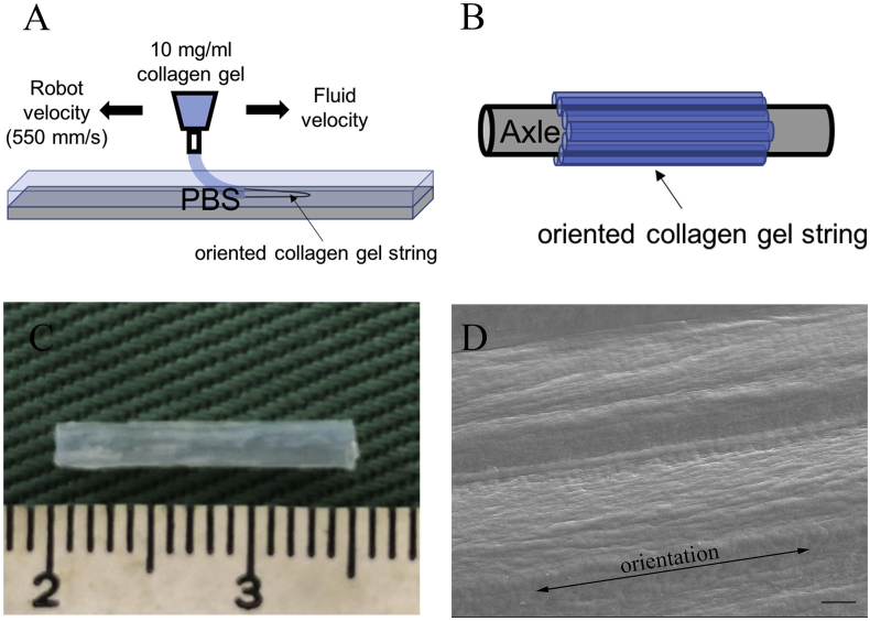
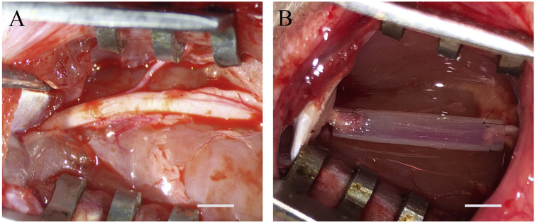
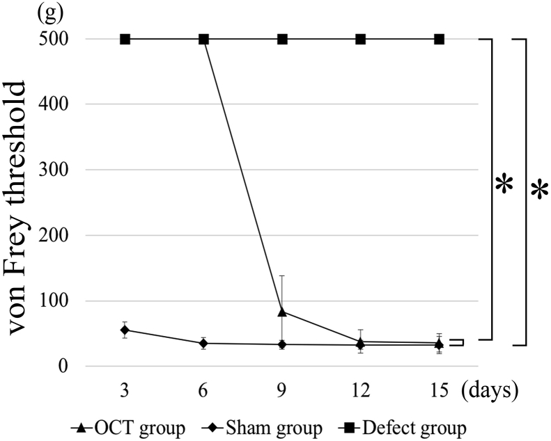
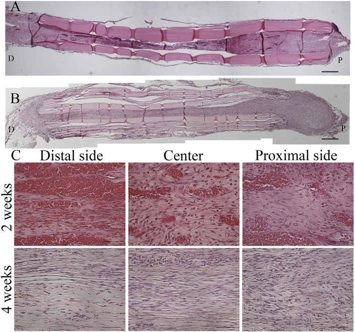
Materials and methods: After a 10-mm long portion of the sciatic nerve of a rat was resected, an OCT was placed in the site of nerve defect. Recovery of the sensory function was evaluated using Von Frey test every 3 days after surgery. The regenerated tissue were histologically and ultrastructurally analyzed 2 and 4 weeks after the surgery.

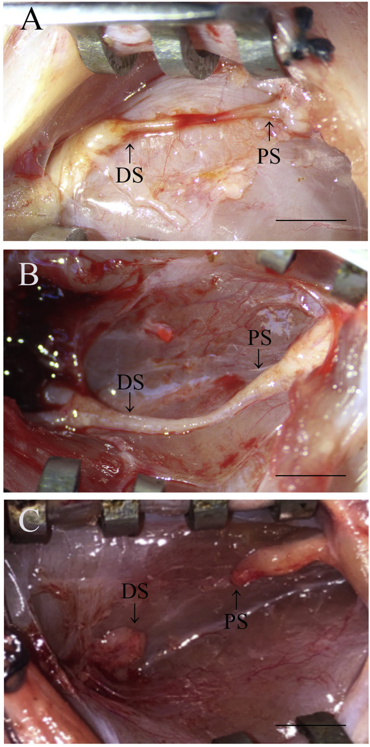
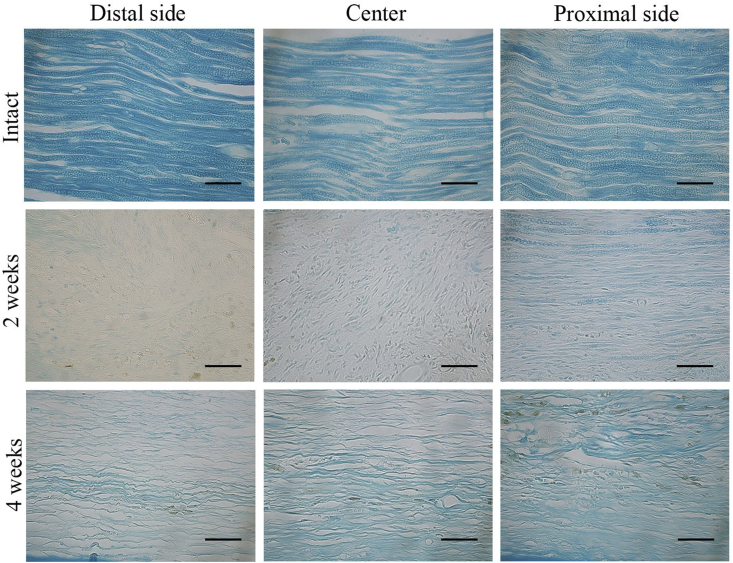
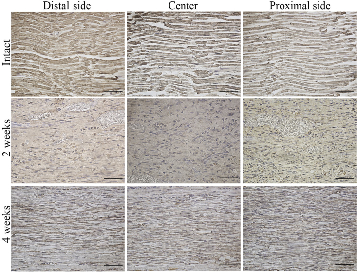
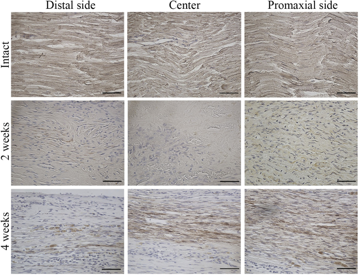
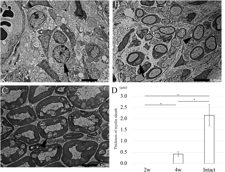
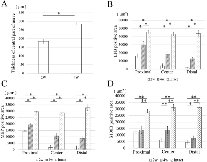
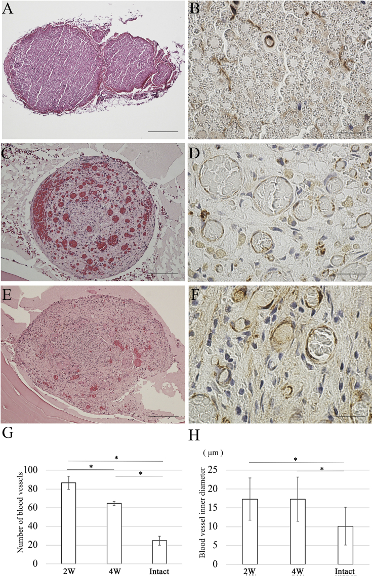
Results: The sensory reflexes of the OCT group were restored to the level of that of the intact group after 15 days. Hematoxylin and eosin staining revealed the cross-linking between the proximal and distal stumps after 2 weeks. After 4 weeks, Luxol Fast Blue and immunohistochemical staining revealed the presence of myelin sheath from the proximal to distal region of the regenerated tissue and S100B staining confirmed the presence of Schwann cells. Interestingly, no myelin sheath was ultrastructurally observed around the regenerated axons at the central region after 2 weeks.

Conclusions: These results suggest that OCTs facilitate the recovery of sensory function. Additionally, the non-myelinated axons contributed to the recovery of the sensory function.

Keywords: Biocompatible materials; H&E, hematoxylin and eosin; IAN, inferior alveolar nerve; LFB, Luxol Fast Blue; MBP, myelin basic protein; Microscopy; Nerve regeneration; OCT, oriented collagen tube; PBS, phosphate buffered saline; PODs, postoperative days; Peripheral nerves; SD, standard deviation; Sciatic nerve; TEM, transmission electron microscopy.


Conflict of interest statement

The authors declare no conflict of interest.

Figures

Fig. 1
Fig. 1
Preparation of oriented collagen tubes (OCTs). (A) Schematic illustration of the robotic deposition of collagen gel in PBS buffer. (B) After the injection of collagen gel strings, they are demineralized and aligned on a polypropylene rod seven times to fabricate a seven-layer seamless tube. (C) An oriented collagen tube (OCT). (D) The scanning electron microscopy image of the OCT indicating the orientation of collagen fibers. Scale bar = 10 μm.
Fig. 2
Fig. 2
OCT implantation. (A) After dissecting the skin of the left thigh, the left sciatic nerve is exposed. (B) A 10-mm long portion of the left sciatic nerve is resected. The OCT is indwelled, and the nerve stump and the OCT are suture at 2 places each with 10-0 nylon thread. Scale bar = 3 mm.
Fig. 3
Fig. 3
Von Frey test. Von Frey test performs every 3 days after surgery. There are significant differences between the sham group and the defect group and between the OCT group and the defect group at postoperative day 15 (P < 0.01).
Fig. 4
Fig. 4
Gross observation. (A) Image of the sciatic nerve 2 weeks after OCT implantation. (B) Image of the sciatic nerve 4 weeks after OCT implantation. (C) Image of the sciatic nerve in the defect group. PS: Proximal stump; DS: Distal stump. Scale bars = 5 mm.
Fig. 5
Fig. 5
Histological analysis by H&E staining in the OCT group. (A) Longitudinal image of the regenerated tissues 2 weeks after OCT implantation (B) and 4 weeks after OCT implantation. (C) High-magnification H&E-stained longitudinal images 2 and 4 weeks after surgery. P: Proximal; D: Distal. A, B Scale bars = 500 μm. C Scale bars = 50 μm.
Fig. 6
Fig. 6
Histological analysis by using Luxol fast blue (LFB) staining. The longitudinal sections showing the proximal region, central region, and distal region of the regenerated tissue from the intact group and the OCT group (2 weeks and 4 weeks after implantation). Scale bars = 50 μm.
Fig. 7
Fig. 7
Myelin basic protein (MBP) expression in the regenerated peripheral nerve. Longitudinal section showing the proximal, central, and distal regions of the regenerated tissue from the intact group and the OCT group (2 weeks and 4 weeks after implantation). Scale bars = 50 μm.
Fig. 8
Fig. 8
S100B protein expression in the regenerated peripheral nerve. Longitudinal sections showing the proximal, central, and distal regions of the regenerated tissue from the intact group and the OCT group (2 weeks and 4 weeks after implantation). Scale bars = 50 μm.
Fig. 9
Fig. 9
Transmission electron microscopy images of cross-sections of a regenerated peripheral nerve. (A) 2 weeks after OCT implantation, the nuclei of Schwann cells surround by unmyelinated axons are observed (arrow). (B) 4 weeks after OCT implantation, myelinated axons are observed (arrowhead). (C) Images of intact tissue. (D) The thickness of the myelin sheath as measured using Image-J soft. Ten myelin sheaths are selected from the central region of the regenerated peripheral nerve. The thickness in the OCT group is significantly lower than that in the intact group (P < 0.01).
Fig. 10
Fig. 10
Results of statistical analyses corresponding toFig. 5,Fig. 6,Fig. 7, andFig. 8. (A) The thickness of regenerated tissue at the central region at the 4-week time point is significantly higher than that at the 2-week time point (P < 0.01). (B) At the 4-week time point, the LFB-positive area from the proximal to distal region is smaller in the OCT group than that in the intact tissues (P < 0.01). (C) At the 4-week time point, the MBP-positive area in the proximal and distal region is smaller than that of the intact tissues. (P < 0.01) (D) The S100B positive area in the proximal and the distal regions is smaller than that in the intact tissue at the 4-week time point (*P < 0.05; **P < 0.01).
Fig. 11
Fig. 11
Evaluation of angiogenesis. Sections of the central region of the regenerated tissues are subjected to H&E staining and immunostaining using anti-CD34 antibodies to detect endothelial cells in the regenerated peripheral nerve. (A) H&E staining image of central region of the regenerated tissue in intact group. (B) Immunostaining using anti-CD34 antibodies: image of the central region of the regenerated tissue in the intact group. (C) H&E staining: image of central region of the regenerated tissue 2 weeks after OCT implantation. (D) Immunostaining using anti-CD34 antibodies: image of the central region of the regenerated tissue 2 weeks after OCT implantation. (E) H&E staining: image of the central region of the regenerated tissue 4 weeks after OCT implantation. (F) Immunostaining using anti-CD34 antibodies: image of the central region of the regenerated tissue 4 weeks after OCT implantation. (G) The number of blood vessels is higher in the OCT group than in the intact group (P < 0.01). (H) The inner diameter of newly formed blood vessels in the OCT group is significantly thicker than the normal thickness (P < 0.01). A, E Scale bars = 200 μm. C Scale bars = 100 μm. B, D, F Scale bars = 20 μm.

References

    1. Faroni A., Mobasseri S.A., Kingham P.J., Reid A.J. Peripheral nerve regeneration: experimental strategies and future perspectives. Adv Drug Deliv Rev. 2015;82–83:160–167. - PubMed
    1. Raso V.V., Barbieri C.H., Mazzer N., Fasan V.S. Can therapeutic ultrasound influence the regeneration of peripheral nerves? J Neurosci Methods. 2005;142(2):185–192. - PubMed
    1. Matsuyama T., Mackay M., Midha R. Peripheral nerve repair and grafting techniques: a review. Neurol Med Chir (Tokyo) 2000;40(4):187–199. - PubMed
    1. Jiang B., Zhang P., Zhang D., Fu Z., Yin X., Zhang H. Study on small gap sleeve bridging peripheral nerve injury. Artif Cells Blood Substit Immobil Biotechnol. 2006;34(1):55–74. - PubMed
    1. Oprych K.M., Whitby R.L., Mikhalovsky S.V., Tomlins P., Adu J. Repairing peripheral nerves: is there a role for carbon nanotubes. Adv Healthc Mater. 2016;5(11):1253–1271. - PubMed

LinkOut - more resources