

Miswiring of Frontostriatal Projections in Schizophrenia
- PMID: 31990358
- PMCID: PMC7342176
- DOI: 10.1093/schbul/sbz129
Miswiring of Frontostriatal Projections in Schizophrenia
Abstract
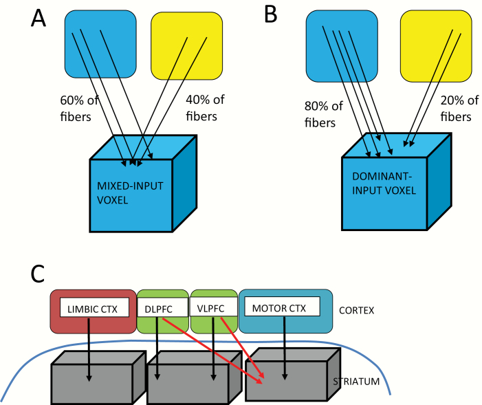
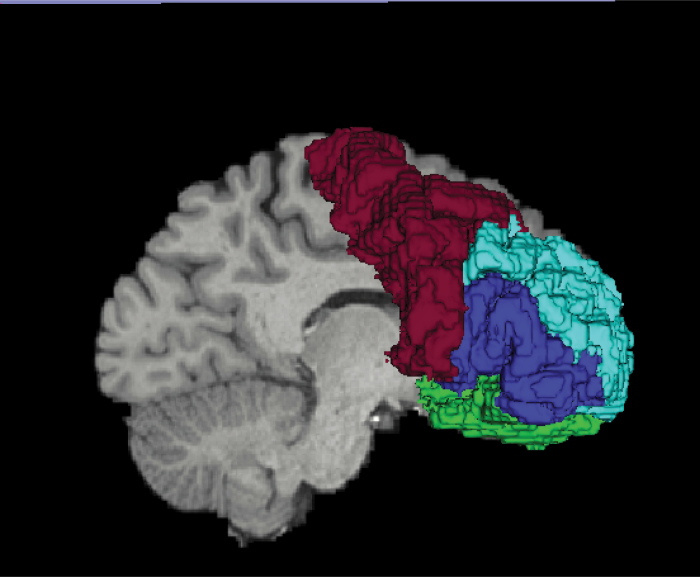
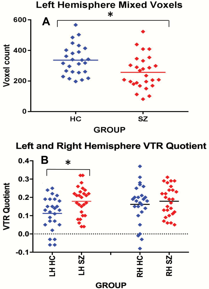
We investigated brain wiring in chronic schizophrenia and healthy controls in frontostriatal circuits using diffusion magnetic resonance imaging tractography in a novel way. We extracted diffusion streamlines in 27 chronic schizophrenia and 26 healthy controls connecting 4 frontal subregions to the striatum. We labeled the projection zone striatal surface voxels into 2 subtypes: dominant-input from a single cortical subregion, and, functionally integrative, with mixed-input from diverse cortical subregions. We showed: 1) a group difference for total striatal surface voxel number (P = .045) driven by fewer mixed-input voxels in the left (P = .007), but not right, hemisphere; 2) a group by hemisphere interaction for the ratio quotient between voxel subtypes (P = .04) with a left (P = .006), but not right, hemisphere increase in schizophrenia, also reflecting fewer mixed-input voxels; and 3) fewer mixed-input voxel counts in schizophrenia (P = .045) driven by differences in left hemisphere limbic (P = .007) and associative (P = .01), but not sensorimotor, striatum. These results demonstrate a less integrative pattern of frontostriatal structural connectivity in chronic schizophrenia. A diminished integrative pattern yields a less complex input pattern to the striatum from the cortex with less circuit integration at the level of the striatum. Further, as brain wiring occurs during early development, aberrant brain wiring could serve as a developmental biomarker for schizophrenia.
Keywords: brain wiring; diffusion magnetic resonance imaging; prefrontal cortex; schizophrenia; striatum; tractography.
Published by Oxford University Press on behalf of the Maryland Psychiatric Research Center 2020.
Figures
References
-
- Alexander GE, Crutcher MD. Functional architecture of basal ganglia circuits: neural substrates of parallel processing. Trends Neurosci. 1990;13(7):266–271. - PubMed
-
- Alexander GE, DeLong MR, Strick PL. Parallel organization of functionally segregated circuits linking basal ganglia and cortex. Annu Rev Neurosci. 1986;9:357–381. - PubMed
-
- Manoach DS, Gollub RL, Benson ES, et al. Schizophrenic subjects show aberrant fMRI activation of dorsolateral prefrontal cortex and basal ganglia during working memory performance. Biol Psychiatry. 2000;48(2):99–109. - PubMed
-
- Voorn P, Vanderschuren LJ, Groenewegen HJ, Robbins TW, Pennartz CM. Putting a spin on the dorsal-ventral divide of the striatum. Trends Neurosci. 2004;27(8):468–474. - PubMed
-
- Bhatia KP, Marsden CD. The behavioural and motor consequences of focal lesions of the basal ganglia in man. Brain. 1994;117(Pt 4):859–876. - PubMed
Publication types
MeSH terms
Grants and funding
LinkOut - more resources
Full Text Sources
Medical
