. 2020 Feb 27;5(4):e135700.
doi: 10.1172/jci.insight.135700.

Metalloproteinase PAPP-A regulation of IGF-1 contributes to polycystic kidney disease pathogenesis

Affiliations

Metalloproteinase PAPP-A regulation of IGF-1 contributes to polycystic kidney disease pathogenesis

Sonu Kashyap et al. JCI Insight. .

Abstract

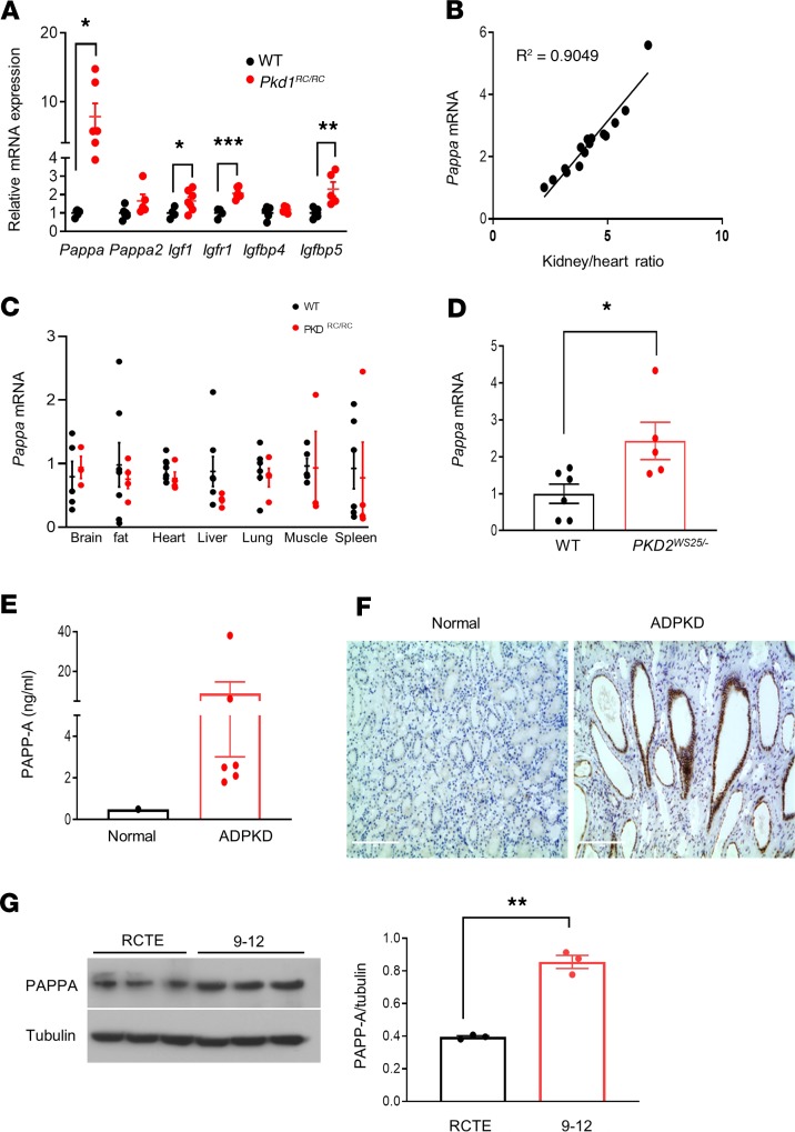
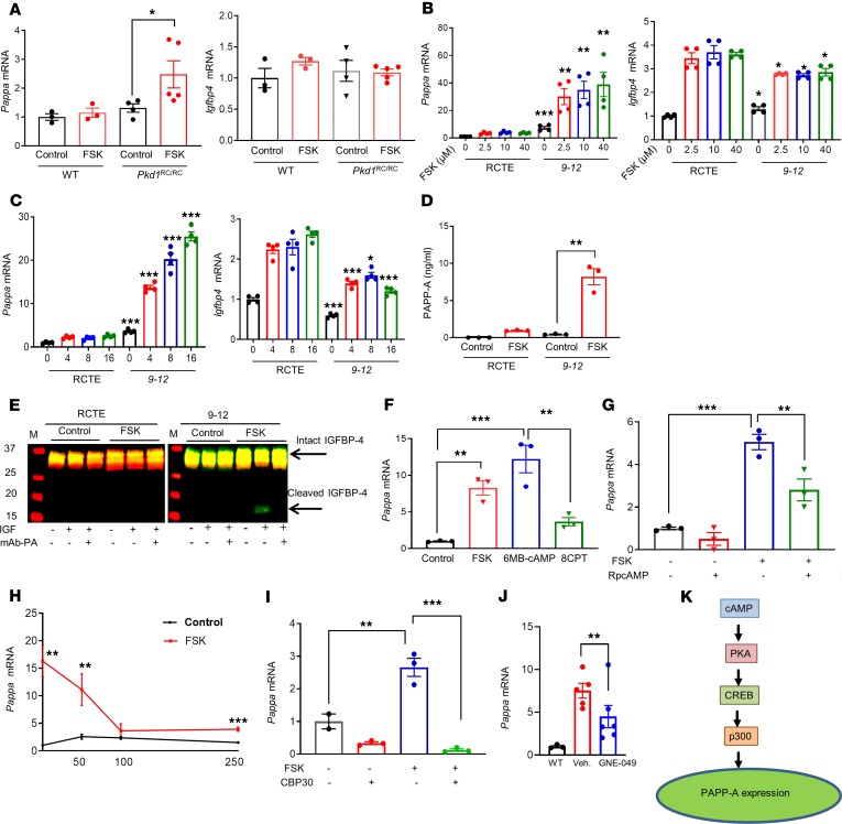
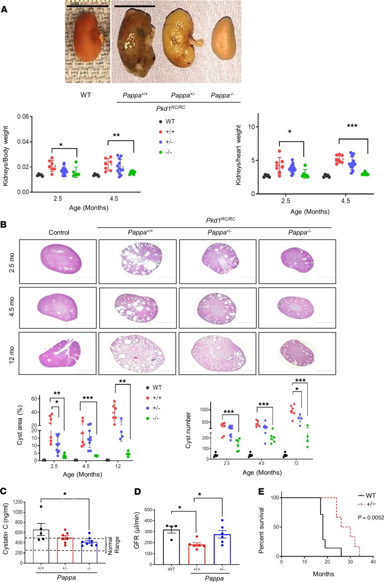
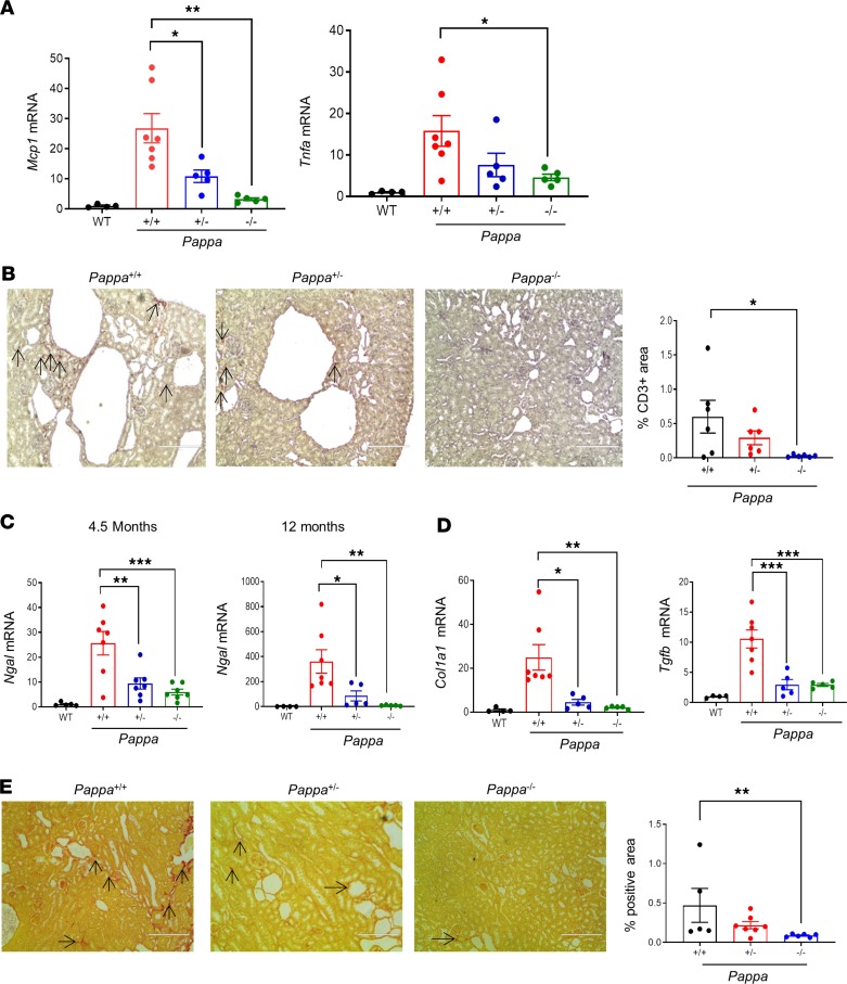
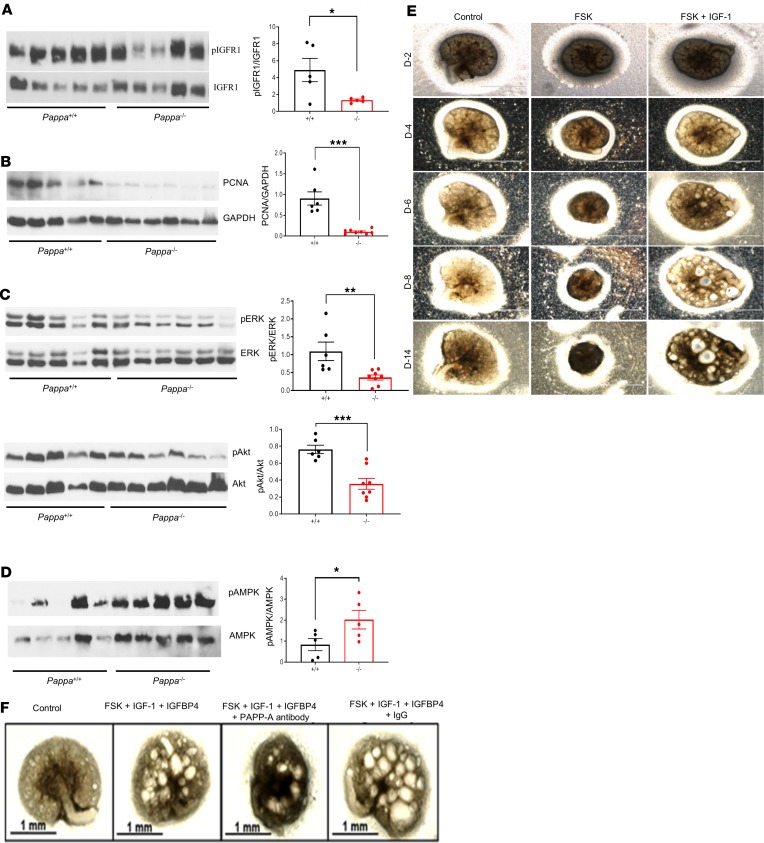
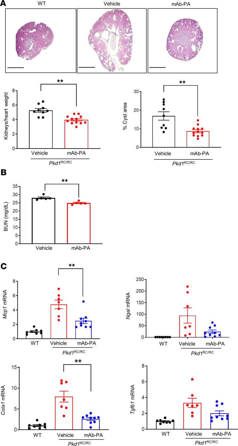
Autosomal dominant polycystic kidney disease (ADPKD) is the most common genetic cause of end-stage renal disease (ESRD). The treatment options for ADPKD are limited. We observed an upregulation in several IGF-1 pathway genes in the kidney of Pkd1RC/RC mice, a model of ADPKD. Pregnancy-associated plasma protein A (PAPP-A), a metalloproteinase that cleaves inhibitory IGF binding proteins (IGFBPs), increasing the local bioactivity of IGF-1, was highly induced in the kidney of ADPKD mice. PAPP-A levels were high in cystic fluid and kidneys of humans with ADPKD. Our studies further showed that PAPP-A transcription in ADPKD was mainly regulated through the cAMP/CREB/CBP/p300 pathway. Pappa deficiency effectively inhibited the development of cysts in the Pkd1RC/RC mice. The role of PAPP-A in cystic disease appears to be regulation of the IGF-1 pathway and cellular proliferation in the kidney. Finally, preclinical studies demonstrated that treatment with a monoclonal antibody that blocks the proteolytic activity of PAPP-A against IGFBP4 ameliorated ADPKD cystic disease in vivo in Pkd1RC/RC mice and ex vivo in embryonic kidneys. These data indicated that the PAPP-A/IGF-1 pathway plays an important role in the growth and expansion of cysts in ADPKD. Our findings introduce a therapeutic strategy for ADPKD that involves the inhibition of PAPP-A.

Keywords: Molecular pathology; Nephrology.


Conflict of interest statement

Conflict of interest: ENC has applied for a US patent on materials and methods for treating polycystic kidney disease (application 62/822,602).

Figures

Figure 1
Figure 1. Upregulation of PAPP-A is a common feature in experimental and human ADPKD.
(A) Relative mRNA expression of IGF-1 pathway components in kidneys of 7.5-month-old C57BL/6J (n = 46) and Pkd1RC/RC mice (n = 5–7). PCR data are expressed relative to Gapdh. (B) Correlation between kidney size (total kidney weight relative to heart weight) and renal Pappa mRNA expression in Pkd1RC/RC mice (n = 15). (C) Pappa mRNA levels in various tissues of WT (n = 3–5) and Pkd1RC/RC mice (n = 4–6). (D) Pappa mRNA levels in WT (n = 6) and Pkd2WS25/– (n = 5) mouse kidneys (16 weeks old). (E) ELISA analysis of PAPP-A protein levels in human ADPKD cystic fluid (n = 6) compared with normal serum reference. (F) Immunolocalization of PAPP-A in normal and ADPKD human kidneys. (G) Western blot analysis of PAPP-A protein levels in normal human RCTE and ADPKD cystic epithelial cells (9-12); graph shows quantification relative to tubulin. Scale bars: 200 μm. Data are expressed as mean ± SEM. *P < 0.05, **P < 0.01, ***P < 0.001 by 2-tailed (for Igf1, 1-tailed) Student’s t test.
Figure 2
Figure 2. Molecular pathways involved in regulation of PAPP-A expression.
(A) Pappa and Igfbp4 mRNA levels in kidneys of Pkd1RC/RC (n = 4–5) and WT mice (n = 3) treated with vehicle (5% DMSO) or 5 mg/kg FSK for 24 hours at 4 weeks of age. (B and C) Pappa and Igfbp4 mRNA levels in RCTE and PKD cystic epithelial cells (9-12) treated with (B) increasing doses of FSK for 4 hours and (C) 10 μM FSK for various time intervals. (D) ELISA analysis of PAPPA levels in cell-free conditioned media of RCTE and 9-12 cells treated with 10 μM FSK or vehicle for 72 hours. (E) Proteolytic assay of PAPP-A–mediated IGFBP4 using cell-free conditioned media of RCTE and 9-12 cells treated with FSK or vehicle for 72 hours. Conditioned medium was incubated for 72 hours at 37°C with IGFBP4 without (−) or with (+) precomplexing to IGF, and without (−) or with (+) the addition of inhibitory mAb-PA 1/41 antibody. Arrows indicate intact and cleaved IGFBP4 bands. (FI) Pappa mRNA expression in 9-12 cells treated with: (F) vehicle control (0.1% DMSO), 10 μM FSK, a selective activator of PKA (6-MB-cAMP, 200 μM) or Epac (8CPT2OMe, 30 μM) for 16 hours; (G) 10 μM FSK in the presence or absence of a competitive antagonist of cAMP (RpcAMP, 100 μM) for 16 hours; (H) 10 μM FSK in the presence or absence of the indicated doses of KG-501, which blocks cAMP-induction of CREB for 16 hours; (I) a selective CBP/p300 bromodomain inhibitor (CBP30, 10 μM) for 24 hours followed by 10 μM FSK for 16 hours. (J) Pappa mRNA expression levels in WT and Pkd1RC/RC treated with the CBP inhibitor GNE-049 (30 mg/kg, n = 5) or vehicle (Veh., n = 5) twice a day for 3 days orally. (K) Schematic representation of cAMP-induced PAPP-A expression. Data are mean ± SEM. *P < 0.05, **P < 0.01, ***P < 0.001 by 2-tailed Student’s t test or Mann Whitney U test, or 1-way ANOVA followed by Tukey’s post hoc test (for B and C, comparison of 9-12 cells is at their respective dose or time with normal cells).
Figure 3
Figure 3. Genetic deletion of Pappa ameliorates ADPKD.
(A and B) Pkd1RC/RC mice that are Pappa+/+, Pappa+/–, or Pappa–/–: (A) Representative gross kidney images at 12 month age, and graphs of kidney/body weight and kidney/heart weight at different ages (n = 5–15). Scale bars: 1 cm. (B) H&E kidney section photomicrographs and graphs of cystic area and cyst number compared with WT mice at different ages (n = 4–10). (C) Serum cystatin C levels in Pkd1RC/RC Pappa mutant mice at 12 months old age (n = 5–8). (D) GFR measurements in WT (n = 4), Pkd1RC/RC Pappa+/+ (n = 6), and Pkd1RC/RC Pappa+/– mice (n = 6) at 11–13 month age. (E) Survival curve of Pkd1RC/RC Pappa mutant mice. Pkd1RC/RC Pappa+/+ mice, n = 7; Pkd1RC/RC Pappa+/– mice, n = 6. Data are presented as mean ± SEM. *P < 0.05, **P < 0.01, ***P < 0.001 by 1-way ANOVA followed by Tukey’s post hoc test for parametric or Kruskal-Wallis test followed by Dunn’s post hoc test for nonparametric distribution or 2-tailed Student’s t test.
Figure 4
Figure 4. Genetic deletion of Pappa reduces renal inflammation, injury, and fibrosis in ADPKD mice.
(A) Renal Mcp1 and Tnfα mRNA expression in WT and Pkd1RC/RC Pappa mutants (n = 4–7) at 12 months of age. (B) Representative photomicrographs showing anti-CD3 immunostaining in kidney sections from 4.5-month-old Pkd1RC/RC Pappa mutant mice, and graph showing quantification of positively stained area (n = 6/group). (C) Renal Ngal mRNA expression in WT and Pkd1RC/RC Pappa mutants at 4.5 and 12 months of age (n = 4–7). (D) Col1a 1 and Tgfb mRNA expression in WT and Pkd1RC/RC Pappa mutants (n = 4–7). (E) Representative photomicrographs of Sirius red staining in kidneys of 4.5-month-old Pkd1RC/RC Pappa mutant mice, and graph showing quantification of positively stained area (n = 5–7). Data are presented as mean ± SEM. *P < 0.05, **P < 0.01 by 1-way ANOVA and then Dunnett’s post hoc test for parametric or Kruskal-Wallis test followed by Dunn’s post hoc test for nonparametric distribution. Two-tailed Mann Whitney U test was used for E. Original magnification, ×200; arrows define the positively stained area.
Figure 5
Figure 5. The role of PAPP-A in the pathogenesis of ADPKD.
Western blot analysis of (A) p-IGFR1/IGFR1 (B) PCNA, (C) p-ERK/ERK and p-Akt/Akt, and (D) p-AMPK/AMPK in kidney tissues of 2.5- or 4.5-month-old Pkd1RC/RC Pappa+/+ and Pkd1RC/RC Pappa–/– mice. Graphs show quantitative analysis of bands by densitometry. (E) Photomicrographs showing that IGF-1 supports cystic growth in a metanephric model of cystogenesis. Day 13.5 embryonic kidneys from WT mice were stimulated with FSK (10 μM) alone (no growth hormone added) or with FSK in the presence of IGF-1 (100 ng/mL). Scale bars: 1 mm. (F) Embryonic kidneys were treated with FSK (10 μM), IGF-1 (100 ng/ml), and IGFBP4 (26 nM) in the presence or absence of mAb-PA (320 pM). IGF-1 was incubated with IGFBP4 prior to treatment. For metanephric culture, the experiment was repeated twice; n = 3 for each experiment. Data are mean ± SEM. *P < 0.05, **P < 0.01, ***P < 0.001 by 2-tailed Student’s t test (1-tailed for D). Scale bars: 1 mm.
Figure 6
Figure 6. Blockade of PAPP-A function with monoclonal antibody (mAb-PA) decreases cyst formation in ADPKD mice.
Pkd1RC/RC mice were treated with mAb-PA (30 mg/kg) or vehicle once per week for 6 weeks. (A) Representative images of H&E-stained kidney sections and graphs showing kidney size and percent cystic area; age, 7.5 months (n = 9–12). Scale bars: 2000 μm. (B) BUN measurements in vehicle (n = 5) and anti–PAPP-A–treated mice (n = 5). (C) Mcp1, Ngal, Col1a1, and Tgfb mRNA expression in vehicle or mAb-PA–treated Pkd1RC/RC mice (n = 7–9). Data are mean ± SEM. A *P < 0.05, **P < 0.01 by 2-tailed Student’s t test.

References

    1. Tan YC, Blumenfeld J, Rennert H. Autosomal dominant polycystic kidney disease: genetics, mutations and microRNAs. Biochim Biophys Acta. 2011;1812(10):1202–1212. - PubMed
    1. Torres VE, Harris PC, Pirson Y. Autosomal dominant polycystic kidney disease. Lancet. 2007;369(9569):1287–1301. doi: 10.1016/S0140-6736(07)60601-1. - DOI - PubMed
    1. Chebib FT, Torres VE. Autosomal dominant polycystic kidney disease: core curriculum 2016. Am J Kidney Dis. 2016;67(5):792–810. doi: 10.1053/j.ajkd.2015.07.037. - DOI - PMC - PubMed
    1. Bolignano D, Palmer SC, Ruospo M, Zoccali C, Craig JC, Strippoli GFM. Interventions for preventing the progression of autosomal dominant polycystic kidney disease. Cochrane Database Syst Rev. 2015;(7):CD010294. - PMC - PubMed
    1. Grantham JJ, Geiser JL, Evan AP. Cyst formation and growth in autosomal dominant polycystic kidney disease. Kidney Int. 1987;31(5):1145–1152. doi: 10.1038/ki.1987.121. - DOI - PubMed

Publication types

MeSH terms

Substances