. 2020 Feb 27;5(4):e135321.
doi: 10.1172/jci.insight.135321.

Fetal and amniotic fluid iron homeostasis in healthy and complicated murine, macaque, and human pregnancy

Affiliations

Fetal and amniotic fluid iron homeostasis in healthy and complicated murine, macaque, and human pregnancy

Allison L Fisher et al. JCI Insight. .

Abstract

Adequate iron supply during pregnancy is essential for fetal development. However, how fetal or amniotic fluid iron levels are regulated during healthy pregnancy, or pregnancies complicated by intraamniotic infection or inflammation (IAI), is unknown. We evaluated amniotic fluid and fetal iron homeostasis in normal and complicated murine, macaque, and human pregnancy. In mice, fetal iron endowment was affected by maternal iron status, but amniotic fluid iron concentrations changed little during maternal iron deficiency or excess. In murine and macaque models of inflamed pregnancy, the fetus responded to maternal systemic inflammation or IAI by rapidly upregulating hepcidin and lowering iron in fetal blood, without altering amniotic fluid iron. In humans, elevated cord blood hepcidin with accompanying hypoferremia was observed in pregnancies with antenatal exposure to IAI compared with those that were nonexposed. Hepcidin was also elevated in human amniotic fluid from pregnancies with IAI compared with those without IAI, but amniotic fluid iron levels did not differ between the groups. Our studies in mice, macaques, and humans demonstrate that amniotic fluid iron is largely unregulated but that the rapid induction of fetal hepcidin by inflammation and consequent fetal hypoferremia are conserved mechanisms that may be important in fetal host defense.

Keywords: Bacterial infections; Homeostasis; Innate immunity; Reproductive Biology.


Conflict of interest statement

Conflict of interest: TG and EN are shareholders of and scientific advisors for Intrinsic LifeSciences and Silarus Therapeutics and consultants for Ionis Pharmaceuticals, Protagonist, and Vifor. TG is a consultant for Akebia.

Figures

Figure 1
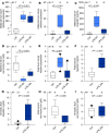
Figure 1. Effect of maternal iron status on fetal and amniotic fluid iron parameters in mice.
Maternal iron deficiency was induced by feeding low-iron diet to WT dams starting on E10.5 until analysis at E18.5. Iron-loaded dams were hepcidin-deficient mice fed standard chow. Both iron-deficient and iron-loaded dams were compared with iron-replete WT dams fed standard chow. (A) Amniotic fluid iron concentrations from iron-replete dams from E12.5 to E18.5. (B and C) Fetal serum and amniotic fluid iron from iron-deficient, iron-replete, and iron-loaded dams on E18.5. (DF) Pearson correlations between maternal serum iron, fetal serum iron, and amniotic fluid iron on E18.5. For D and E, maternal serum iron was correlated to the litter average for fetal serum iron or amniotic fluid iron. Statistical differences between groups were determined by 1-way ANOVA on ranks followed by Dunn’s method for multiple comparisons (as indicated by #). The number of animals is indicated above the box plot for each panel.
Figure 2
Figure 2. Effect of maternal iron status on fetal and amniotic fluid transferrin and transferrin saturation in mice.
Maternal iron deficiency was induced by feeding low-iron diet to WT dams starting on E10.5 until analysis at E18.5. Iron-loaded dams were hepcidin-deficient mice fed standard chow. Both iron-deficient and iron-loaded dams were compared with iron-replete WT dams fed standard chow. (A and B) Transferrin concentrations and TSAT in maternal serum, amniotic fluid, and fetal serum on E18.5 in iron-replete pregnancy. (C and D) Maternal transferrin concentration and TSAT and (EH) fetal serum and amniotic fluid transferrin concentration and TSAT from iron-deficient, iron-replete, and iron-loaded pregnancies on E18.5. Statistical differences between groups were determined by 1-way ANOVA for normally distributed values followed by Holm-Sidak method for multiple comparisons (as indicated by *) or 1-way ANOVA on ranks followed by Dunn’s method for multiple comparisons (as indicated by #). The number of animals is reported above the box plot for each panel.
Figure 3
Figure 3. Effect of maternal systemic inflammation on fetal iron homeostasis in mice.
To induce maternal systemic inflammation during pregnancy, iron-replete WT dams received a single subcutaneous injection of 0.5 μg/g LPS on E15.5 for 6 or 24 hours. (A) Maternal liver serum amyloid A-1 (Saa1) and (B) hepcidin (Hamp) mRNA expression normalized to Hprt. Measurements in maternal serum: (C) hepcidin and (D) iron. Fetal (E) liver hepcidin mRNA expression normalized to Rpl4, (F) serum hepcidin, and (G) serum iron. Amniotic fluid (H) hepcidin and (I) iron. Statistical differences between groups were determined by 1-way ANOVA for normally distributed values followed by Holm-Sidak method for multiple comparisons (as indicated by *), 2-tailed Student’s t test for normally distributed values (as indicated by #), or Mann-Whitney U test (as indicated by &). The number of animals is reported above the box plot for each panel.
Figure 4
Figure 4. Cytokines in maternal plasma, cord blood plasma, and amniotic fluid during IAI in rhesus macaques.
Pregnant rhesus macaques at 130 days gestation received a single intraamniotic injection of LPS (1 mg) for 16 hours or Ureaplasma parvum serovar 1 (1 × 10⁷ CFU) for 3 days. Cytokines TNF-α, MCP-1, IL-1β, and IL-6 were measured in (AD) maternal plasma, (EH) cord blood plasma, and (IL) amniotic fluid at delivery. Statistical differences between groups were determined by 1-way ANOVA for normally distributed values followed by Holm-Sidak method for multiple comparisons (as indicated by *) or 1-way ANOVA on ranks followed by Dunn’s method for multiple comparisons (as indicated by #). A subset of these data was previously reported (30, 44). The number of animals is reported on the x axis of each panel.
Figure 5
Figure 5. Iron parameters in maternal plasma, cord blood plasma, and amniotic fluid during IAI in rhesus macaques.
Pregnant rhesus macaques received a single intraamniotic injection of LPS (1 mg) for 16 hours or Ureaplasma parvum serovar 1 (1 × 10⁷ CFU) for 3 days. (AC) Hepcidin and (DF) iron measurements in maternal plasma, cord blood plasma, and amniotic fluid. The control group included samples taken before injection or after saline injection (there was no difference between the 2 groups). The saline group included only samples at delivery after saline injection. Statistical differences between groups were determined by 1-way ANOVA for normally distributed values followed by Holm-Sidak method for multiple comparisons (as indicated by *) or 1-way ANOVA on ranks followed by Dunn’s method for multiple comparisons (as indicated by #). The number of animals is reported above the box plot for each panel.
Figure 6
Figure 6. Cord blood iron homeostasis in healthy and complicated human pregnancy.
Cord blood from the umbilical vein was sampled at the time of delivery from singleton preterm human fetuses (<34 weeks of gestational age) with or without antenatal exposure to intraamniotic infection. Measurements at time of delivery in cord blood plasma: (A) hepcidin, (B) IL-6, (C) non-heme iron, and (D) transferrin saturation. Statistical differences between groups were determined by 1-way ANOVA on ranks followed by Dunn’s method for multiple comparisons (as indicated by #). The number of samples are reported above the box plot for each figure panel.

Similar articles

Cited by

References

    1. Breuer W, Ronson A, Slotki IN, Abramov A, Hershko C, Cabantchik ZI. The assessment of serum nontransferrin-bound iron in chelation therapy and iron supplementation. Blood. 2000;95(9):2975–2982. doi: 10.1182/blood.V95.9.2975.009k03_2975_2982. - DOI - PubMed
    1. Evans P, Cindrova-Davies T, Muttukrishna S, Burton GJ, Porter J, Jauniaux E. Hepcidin and iron species distribution inside the first-trimester human gestational sac. Mol Hum Reprod. 2011;17(4):227–232. doi: 10.1093/molehr/gaq101. - DOI - PMC - PubMed
    1. Gazzolo D, et al. Non protein bound iron concentrations in amniotic fluid. Clin Biochem. 2005;38(7):674–677. doi: 10.1016/j.clinbiochem.2005.03.010. - DOI - PubMed
    1. Hattori Y, et al. Catalytic ferrous iron in amniotic fluid as a predictive marker of human maternal-fetal disorders. J Clin Biochem Nutr. 2015;56(1):57–63. doi: 10.3164/jcbn.14-82. - DOI - PMC - PubMed
    1. Legge M. Second trimester amniotic fluid ferritin and transferrin levels in normal and anencephalic pregnancies. J Obstet Gynaecol (Lahore) 1983;4(1):14–15. doi: 10.3109/01443618309071216. - DOI

Publication types