A Novel Method of CT Exophthalmometry in Patients With Thyroid Eye Disease
- PMID: 31990744
- PMCID: PMC7004459
- DOI: 10.1097/01.APO.0000617908.29733.84
A Novel Method of CT Exophthalmometry in Patients With Thyroid Eye Disease
Abstract
Purpose: Conventional computed tomography (CT) exophthalmometry requires an intact lateral orbital wall and is therefore not feasible in patients who have undergone any form of lateral orbital wall surgery where the normal bony landmark may be lost or displaced. The purpose of our study is to validate an alternative method of CT exophthalmometry utilizing the posterior clinoid (PC) process as a new reference point that will allow for reproducible comparison of the anterior-posterior globe position in the preoperative and postoperative settings.
Design: Cohort study.
Methods: This is a retrospective study of 48 patients with clinically diagnosed thyroid eye disease who had undergone cross-sectional CT imaging in the pre- or postoperative settings. CT exophthalmometry was performed using both the conventional interzygomatic method and our proposed PC process method on all pre- and postoperative CT imaging by two independent observers. Interobserver variability analysis was performed with intraclass correlation coefficient. Correlation and agreement between the two methods were analyzed with Pearson correlation coefficient and linear regression method. All analyses were conducted at 5% level of significance with Stata MP V14.
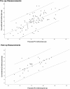
Results: Interobserver variability analysis showed an intraclass correlation coefficient of >0.9 for both interzygomatic and PC methods. There is good correlation between the two different measurements observed in both the pre- and postoperative groups (r = 0.68 and r = 0.72, respectively, P < 0.001). Linear regression showed good agreement between the two different measurements with most of the points lying within the 95% limits.
Conclusions: Our new method agrees well with the conventional method and has the added benefit of being able to reliably assess the anterior-posterior globe position in patients who do not have intact lateral orbital walls after decompressive surgery.
Conflict of interest statement
The authors report no conflicts of interest. The authors alone are responsible for the content and writing of the article.
Figures


References
-
- Nkenke E, Maier T, Benz M, et al. Hertel exophthalmometry versus computed tomography and optical 3D imaging for the determination of the globe position in zygomatic fractures. Int J Oral Maxillofac Surg 2004; 33:125–133. doi:10.1054/ijom.2002.0481. - PubMed
-
- Kashkouli MB, Beigi B, Noorani MM, et al. Hertel exophthalmometry: reliability and interobserver variation. Orbit 2003; 22:239–245. - PubMed
-
- Shan SJ, Douglas RS. The pathophysiology of thyroid eye disease. J Neuroophthalmol 2014; 34:177–185. doi:10.1097/WNO.0000000000000132. - PubMed
-
- Khong JJ, McNab AA, Ebeling PR, et al. Pathogenesis of thyroid eye disease: review and update on molecular mechanisms. Br J Ophthalmol 2016; 100:142–150. doi:10.1136/bjophthalmol-2015-307399. - PubMed

