. 2020 Jan 28;15(1):e0226838.
doi: 10.1371/journal.pone.0226838. eCollection 2020.

Crystal structure of PMGL2 esterase from the hormone-sensitive lipase family with GCSAG motif around the catalytic serine

Affiliations

Crystal structure of PMGL2 esterase from the hormone-sensitive lipase family with GCSAG motif around the catalytic serine

Konstantin M Boyko et al. PLoS One. .

Abstract

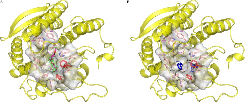
Lipases comprise a large class of hydrolytic enzymes which catalyze the cleavage of the ester bonds in triacylglycerols and find numerous biotechnological applications. Previously, we have cloned the gene coding for a novel esterase PMGL2 from a Siberian permafrost metagenomic DNA library. We have determined the 3D structure of PMGL2 which belongs to the hormone-sensitive lipase (HSL) family and contains a new variant of the active site motif, GCSAG. Similar to many other HSLs, PMGL2 forms dimers in solution and in the crystal. Our results demonstrated that PMGL2 and structurally characterized members of the GTSAG motif subfamily possess a common dimerization interface that significantly differs from that of members of the GDSAG subfamily of known structure. Moreover, PMGL2 had a unique organization of the active site cavity with significantly different topology compared to the other lipolytic enzymes from the HSL family with known structure including the distinct orientation of the active site entrances within the dimer and about four times larger size of the active site cavity. To study the role of the cysteine residue in GCSAG motif of PMGL2, the catalytic properties and structure of its double C173T/C202S mutant were examined and found to be very similar to the wild type protein. The presence of the bound PEG molecule in the active site of the mutant form allowed for precise mapping of the amino acid residues forming the substrate cavity.


Conflict of interest statement

The authors have declared that no competing interests exist.

Figures

Fig 1
Fig 1. Structure of PMGL2 esterase.
(A) Cartoon diagram of the wtPMGL2 subunit. Secondary structure elements are color coded (α-helix—red; β-strand—yellow; loop—green). Characteristic sequence motifs are highlighted as followed: 105HGGAF109(blue), 269GGRD272(magenta), 296FxxGxxHxxF305(grey), 172GCSAG176(cyan) and 137YRLA140(black). CAP-domain is highlighted in orange. The loop 216–237 is depicted in dark green. (B) Comparison of PMGL2 subunit (green, semitransparent) with homologous structure of esterase E25 from metagenomic library from the South China Sea (magenta, PDB entry 4Q05). Orientation of the wtPMGL2 is similar to panel A. Loop 216–237 of wtPMGL2 is highlighted in blue and corresponding loop of 4Q05 –in red.
Fig 2
Fig 2. Multiple sequence alignment of PMGL2 and homologous enzymes with known three-dimensional structure.
The extent of amino acid sequence conservation is depicted in grades of blue. PMGL2 secondary structure elements are indicated at the top. Black circles mark amino acid residues belonging to the catalytic triad.
Fig 3
Fig 3. Comparison of the PMGL2 dimer with the dimers of homologous enzymes.
(A) Cartoon representation of the mPMGL2 dimer. Enzyme subunits are highlighted in different colors. CAP-domains and β8 strands are colored in magenta and blue, respectively. Magnesium ions are shown as red spheres. (B) Surface representation of the mPMGL2 dimer. Hereinafter orientation of the molecules and color scheme are the same as on the panel A. PEG molecules bound in the active site cavity are depicted in pink. Entrances to the active site are marked with red ellipses, reflecting their approximate size. (C) Cartoon representation of the 4Q05 dimer. (D) Surface representation of the 4Q05 dimer. The second entrance to the active site on the side of the molecule is marked with an arrow. (E) Cartoon representation of the 3K6K dimer. (F) Surface representation of the 3K6K dimer. The second entrance to the active site is not marked as it is situated on the opposite side of the molecule. Note that in this case, β8 strands lie near in the same plane (are antiparallel).
Fig 4
Fig 4. mPMGL2 active site entrance.
(A) Amino acid residues of the PMGL2 that form the entrance are shown as semi-transparent grey surface and pink sticks and labeled in magenta. PEG molecule is shown in magenta together with its omit Fo-Fc map at 3σ level (green). Major residues restricting the active site cavity are shown and labeled in red. (B) Residues restricting active site cavity of 4Q05 (black) and 3K6K (blue) are superposed on PMGL2 structure from panel A.
Fig 5
Fig 5. Residues of special interest in the PMGL2 active site.
(A) Two cysteine residues in the wtPMGL2 active site with an appropriate omit Fo-Fc map at 3σ level (gray). Catalytic triad is also depicted. For clarity the rest of molecule is show as a semitransparent green cartoon. (B) The same region in the mPMGL2 active site containing mutations C173T and C202S with an appropriate omit Fo-Fc map at 3σ level. The bound PEG molecule is shown in magenta. The orientation and the color scheme are the same as on panel A.

Similar articles

Cited by

References

    1. Kumar L, Awasthi G, Singh B. Extremophiles: a novel source of industrially important enzymes. Biotechnology. 2011;10(2):121–35.
    1. Karan R, Capes MD, DasSarma S. Function and biotechnology of extremophilic enzymes in low water activity. Aquatic Biosyst. 2012;8(1):1. - PMC - PubMed
    1. Elleuche S, Schröder C, Sahm K, Antranikian G. Extremozymes—biocatalysts with unique properties from extremophilic microorganisms. Curr Opin Biotechnol. 2014;29:116–23. 10.1016/j.copbio.2014.04.003 - DOI - PubMed
    1. Margesin R, Neuner G, Storey K. Cold-loving microbes, plants, and animals—fundamental and applied aspects. Naturwissenschaften. 2007;94(2):77–99. 10.1007/s00114-006-0162-6 - DOI - PubMed
    1. Simon C, Daniel R. Metagenomic analyses: past and future trends. Appl Environ Microbiol. 2011;77(4):1153–61. 10.1128/AEM.02345-10 - DOI - PMC - PubMed

Publication types

MeSH terms