. 2020 Jan 28;15(1):e0221914.
doi: 10.1371/journal.pone.0221914. eCollection 2020.

Embryonic and foetal expression patterns of the ciliopathy gene CEP164

Affiliations

Embryonic and foetal expression patterns of the ciliopathy gene CEP164

L A Devlin et al. PLoS One. .

Abstract

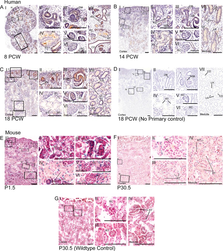
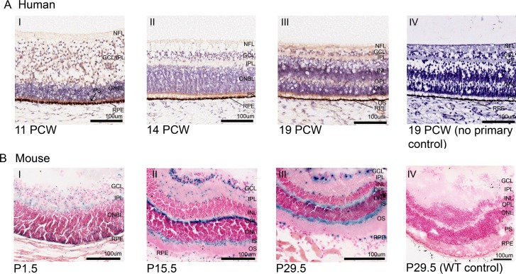
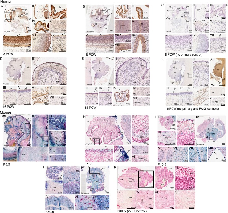
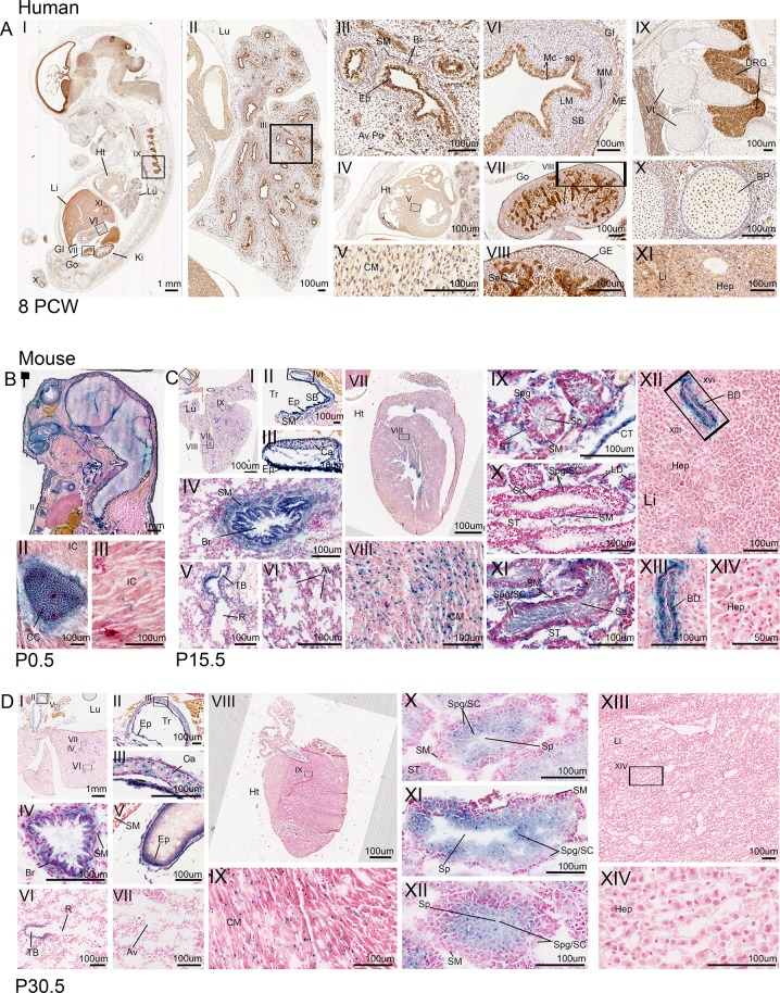
Nephronophthisis-related ciliopathies (NPHP-RC) are a group of inherited genetic disorders that share a defect in the formation, maintenance or functioning of the primary cilium complex, causing progressive cystic kidney disease and other clinical manifestations. Mutations in centrosomal protein 164 kDa (CEP164), also known as NPHP15, have been identified as a cause of NPHP-RC. Here we have utilised the MRC-Wellcome Trust Human Developmental Biology Resource (HDBR) to perform immunohistochemistry studies on human embryonic and foetal tissues to determine the expression patterns of CEP164 during development. Notably expression is widespread, yet defined, in multiple organs including the kidney, retina and cerebellum. Murine studies demonstrated an almost identical Cep164 expression pattern. Taken together, these data support a conserved role for CEP164 throughout the development of numerous organs, which, we suggest, accounts for the multi-system disease phenotype of CEP164-mediated NPHP-RC.


Conflict of interest statement

The authors have declared that no competing interests exist.

Figures

Fig 1
Fig 1. Expression of CEP164 throughout human and murine renal development.
Human renal development (A-D), 8 PCW (A), 14 PCW (B), 18 PCW (C) respectively and 18 PCW no primary control (D). In the developing human kidney, CEP164 expression is seen in the apical membrane of the metanephric renal vesicles (A.II, B.II), s-shaped bodies (B.II, C.I, C.III) and developing renal tubules (A.IV.VII, B.IV.VII). From 14 PCW CEP164 expression is seen at the apical membrane of all developing tubular segments including the distal and proximal tubules (B.IV, C.IV) and loop of Henle segments (B.VII, C.VII). CEP164 expression is seen in the cells of the ureteric bud (A.III, B.III, C.II), and the subsequent collecting duct at both the apical and basal membrane (A.VII, B.VII, C.VII). CEP164 expression is seen in the glomerulus of the developing immature renal corpuscle (A.V, B.V, C.V), and weakly in the matured renal corpuscle (A.VI, B.VI, C.VI). No primary controls demonstrate no background DAB staining, as represented by 18 PCW time-point (D.I-VII). Murine postnatal renal development (E-G), P1.5 (E), P30.5 (F) and P30.5 WT control (G). In the murine kidney Cep164 expression is present in the developing renal vesicles (E.I.II) and ubiquitously throughout subsequent nephron renal tubules (E.IV, F.I.III.IV). Cep164 expression is seen in the glomerulus of the developing renal corpuscle (E.V), but expression seems to be lost with maturity (E.VI, F.II). No endogenous beta galactosidase staining is present in the murine kidney, as shown by the representative image WT control at P30.5 (G). All scale bars represent 100 μm. Cap mesenchyme (CM), collecting duct (CD), distal tubule (DT), loop of Henle (LH), postnatal day (P), post conception weeks (PCW), proximal tubule (PT), renal corpuscle (RC), renal tubule (RT), renal vesicle (RV), s-shaped body (SSB), ureteric bud (UB).
Fig 2
Fig 2. CEP164 expression in the developing human and murine retina.
Human Retina (A), 11 PCW (A.I), 14 PCW (A.II), 19 PCW (A.III) and 19 PCW no primary control (A.IV). In the developing human retina, weak CEP164 expression is seen in the nerve fibre layer (NFL) and ganglion cell layer/inner plexiform layer (GCL/IPL) (A.I) and strong expression in outer neuroblastic (ONBL) photoreceptor precursors (black arrow) (A.I). By 14 PCW, CEP164 expression is seen in the developed inner plexiform layer (IPL) and the developing photoreceptor layers (A.II). At 19 PCW, CEP164 expression is seen in the nerve fibre layer (NFL), inner plexiform layer (IPL), outer plexiform layer (OPL) and photoreceptor layer, with enhancement in the inner photoreceptor segments (IS) (A.III). No background staining is present in the no primary controls (A.IV). Murine Retina (B), P1.5 (B.I), P15.5 (B.II), P29.5 (B.III) and P29.5 WT control (B.IV). In the developing murine retina, Cep164 expression is seen in the inner plexiform layer (IPL), ganglion cell layer (GCL) and outer neuroblastic layer (ONBL) (B.I). At P15.5, Cep164 expression is seen in the ganglion cell layer (GCL), the outer plexiform layer (OPL) and inner segment (IS) of the photoreceptor layer (B.II). There is also punctate expression in the inner plexiform layer (IPL) and edges of the nuclear cell layers (B.II). Retinal pigment epithelium (RPE) also shows Cep164 expression (B.I, B.II). This murine retinal expression patterning is maintained once retinal layers have been formed (B.III). Controls demonstrate no endogenous beta galactosidase activity in the murine retina (B.IV). Ganglion cell layer (GCL), inner nuclear layer (INL), inner plexiform layer (IPL), inner segment (IS), nerve fibre layer (NFL), outer neuroblastic cell layer (ONBL), outer nuclear layer (ONL), outer plexiform layer (OPL), outer segment (OS), photoreceptor segment layer (PS), retinal pigment epithelium (RPE).
Fig 3
Fig 3. CEP164 expression in the developing human and murine brain, focusing on the hindbrain.
Developing human brain (A-F), 8 PCW (A-B), 8 PCW no primary control (C), 16 PCW hindbrain (D),18 PCW hindbrain (E) and 16 PCW hindbrain no primary control (F). In the developing human brain at 8 PCW, CEP164 expression is seen in the neuroepithelium surrounding the telencephalon (A.I.II.VI, B.I.VI), diencephalon (A.I), mesencephalon (A.I), metencephalon (A.I.V) and myencephalon (A.I.VIII, B.I.IV.V), with defined expression in apical neuroepithelium cells lining the brain ventricles (A.I.VI.VIII, B.I.VI.VII.VIII) demonstrated by black arrows. Defined expression is present in the layers of the telencephalon (A.VI, B.VI). There is defined expression of CEP164 in the human brain midline (B.IV.V) and the nasal epithelium (B.IX). At 8PCW, the choroid plexus shows strong CEP164 expression in the ependymal cells (A.II.III.IV, B.II.III), with seemingly weaker expression in the choroid plexus pia matter (A.IV, B.II.III). The ependymal expression is maintained at 16 PCW and 18 PCW (D.I.V, E.I.V). CEP164 expression is seen in the migrating molecular cell layer of the developing cerebellum (D.I.II), which seems to be lost with molecular cell layer maturation (E.I.II). In the human hindbrain the apical membrane of the pons and the cerebellum demonstrate defined CEP164 expression (D.III,IV), which appears to be lost with maturation (E.III.IV). Weak CEP164 expression is seen in the human medulla oblongata (D.VI, E.VI) but not in the white matter (D.VII, E.VII). Developing murine brain (G-K), P0.5 brain (G) P0.5 hindbrain (H), P15.5 (I),P30.5 (J) and P30.5 WT control (K).Cep164 expression is widespread throughout the developing murine brain (P0.5-P30.5) (G.I, I.IV, J.IV) with expression in the cortex, striatum and cerebrum of the maturing cerebral cortex (G.I.VI, I.IV.V, J.IV.V) expression in the midbrain (G.I, I.IV.VII, J.IV.VI) and thalamus (I.IV.VI). Cep164 expression is strong and defined in the ventricular neuroepithelium (I.VII.VIII). The murine choroid plexus demonstrates strong Cep164 expression, however it is not defined to a specific cell type (G.III, H.IV). In the murine hindbrain, at P0.5, Cep164 is expressed in the migrating molecular layer of the cerebellum (G.II.IV, HI.II), this is maintained with expression also in the molecular layer, ganglion cell layer and Purkinje cell layer at P15.5 and P30.5(I.I.II,J.I.II). Cep164 expression is seen in the in the murine pons (G.VII, H.V, J. VII) and medulla oblongata (G.VIII), and weakly in the cerebellar white matter (G.IV, H.VI, I.III), which seems to be lost with maturation (J.III). The representative P30.5 control demonstrates no endogenous beta galactosidase staining (K). Aquaduct (Aq), axon tract (AT), cerebellum (Ce), cerebral cortex (CC), cerebral hemisphere (CH), cerebrum (Cr), choroid plexus (CP), cortex (Cor), cortical plate (CorP), diencephalon (Di), ependymal cells (Ep), fourth ventricle (FV), ganglion cell layer (GCL), inner plexiform layer (IPL), intermediate zone (IL), lateral ventricle (LV), marginal layer (MaL), medulla oblongata (MO), mesencephalon (Mes), metencephalon (Met), midbrain (MB), midline (Mi), molecular cell layer (MCL), myencephalon (Mye), nasal epithelium (Na), outer neuroblastic layer (ONBL), pia (P), pons (PO), purkinje cell layer (PCL), retinal pigment epithelium (RPE), striatum (St), sub-ventricular layer (SVL), telencephalon (Tel), thalamus (Th), third ventricle (TV), ventricular layer (VL), ventricular surface (VS), white matter (WM).
Fig 4
Fig 4. CEP164 expression in secondary organs throughout human and murine development.
Human 8 PCW (A). Murine (B-D), P0.5 (B), P15.5 (C), P30.5 (D). In the developing human, lung CEP164 expression is seen in the respiratory epithelial lining of the bronchi and bronchioles, with weaker expression in the smooth muscle and alveoli (AI.II.III). In the developing murine lung, Cep164 is expressed in the respiratory epithelial lining of the bronchi and bronchioles (C.I.IV, D.I.IV), with additional Cep164 expression seen in the respiratory epithelial cells lining the trachea and the tertiary bronchioles (C.I.II.III.V, D.I.II.III.V.VI). Cep164 expression is seen in murine alveoli at P15.5 (C.VI), which seems to be lost with maturity (P30.5) (D.VII). Cep164 expression is seen within the cartilage of the trachea (C.II.III, D.II.III). In both the human (A.IV.V) and murine hearts (C.VII.VIII, D.VIII.IX), expression is seen the developing cardiomyocytes. In the human gastrointestinal tract CEP164 expression is seen in the inner mucosae squamous epithelial cell layer, muscularis mucosae cell layer and the external muscularis cell layer (A.VI). In the developing human gonads, CEP164 expression is seen in the germline epithelium and seminiferous cord (A.VII.VIII). In the developing murine testes, at P15.5, Cep164 expression is seen in the seminiferous tubules, specifically the smooth muscle cells spermatogonia, spermatocytes and most strongly in spermatids (C.IX.X.XI); expression in leydig cells and connective tissue can also be seen (C.X). At P30.5, Cep164 expression is defined to the spermatogonia, spermatocytes and spermatids (D.X.XI.XII). CEP164 expression is seen in the dorsal root ganglia of the human spinal cord, (A.I.IX) with weaker expression also present in the vertebrae primordia (A.I.IX) and bone primordia (A.I.X). Cep164 expression is seen in the murine developing costal cartilage (B.I.II), with weaker expression in intercostal muscle (B.I.III). The human foetal liver demonstrates some evidence of CEP164 expression (A.XI), however this is undetermined due to high endogenous peroxidase activity in the liver. The developing murine liver shows Cep164 expression in epithelial cells lining the hepatic portal veins at P15.5 (C.XII.XIII), this is not seen by P30.5. There is no expression in hepatocytes (C.XIV, D.XIV). Aveoli (Av), alveoli primordia (Av Pri), bile duct (BD), bone primordia (BP), bronchiole (Br), cardiomyocyte (CM), cartilage (Ca), connective tissue (CT), costal cartilage (CC), dorsal root ganglia (DRG), epithelial (Ep), gastrointestinal tract (GI), germline epithelium (GE), gonad (Go), heart (Ht), hepatocytes (Hep), intercostal muscle (IC), kidney (Ki), lamina propria (LM), liver (Li), lung (Lu), muscularis externa (ME), muscularis mucosae (MM), respiratory bronchiole (R), seminiferous cord (SeC), seminiferous tubule (ST), smooth muscle (SM), spermatids (Sp), spermatocytes (SC), spermatogonia (Spg), squamous cell mucosae (Mc-sq), submucosae (SB), trachea (Tr), terminal bronchi (TB), vertebrae (Vt).

References

    1. Chaki M, Airik R, Ghosh AK, Giles RH, Chen R, Slaats GG, et al. Exome capture reveals ZNF423 and CEP164 mutations, linking renal ciliopathies to DNA damage response signaling. Cell. 2012;150(3):533–48. 10.1016/j.cell.2012.06.028 - DOI - PMC - PubMed
    1. Graser S, Stierhof YD, Lavoie SB, Gassner OS, Lamla S, Le Clech M, et al. Cep164, a novel centriole appendage protein required for primary cilium formation. J Cell Biol. 2007;179(2):321–30. 10.1083/jcb.200707181 - DOI - PMC - PubMed
    1. Utsch B, Sayer JA, Attanasio M, Pereira RR, Eccles M, Hennies HC, et al. Identification of the first AHI1 gene mutations in nephronophthisis-associated Joubert syndrome. Pediatr Nephrol. 2006;21(1):32–5. 10.1007/s00467-005-2054-y - DOI - PubMed
    1. Delous M, Baala L, Salomon R, Laclef C, Vierkotten J, Tory K, et al. The ciliary gene RPGRIP1L is mutated in cerebello-oculo-renal syndrome (Joubert syndrome type B) and Meckel syndrome. Nat Genet. 2007;39(7):875–81. 10.1038/ng2039 - DOI - PubMed
    1. Maria BL, Hoang KB, Tusa RJ, Mancuso AA, Hamed LM, Quisling RG, et al. "Joubert syndrome" revisited: key ocular motor signs with magnetic resonance imaging correlation. J Child Neurol. 1997;12(7):423–30. 10.1177/088307389701200703 - DOI - PubMed

Publication types