. 2020 Mar 4;28(3):901-913.
doi: 10.1016/j.ymthe.2020.01.012. Epub 2020 Jan 15.

Targeting SOX2 Protein with Peptide Aptamers for Therapeutic Gains against Esophageal Squamous Cell Carcinoma

Affiliations

Targeting SOX2 Protein with Peptide Aptamers for Therapeutic Gains against Esophageal Squamous Cell Carcinoma

Kuancan Liu et al. Mol Ther. .

Abstract

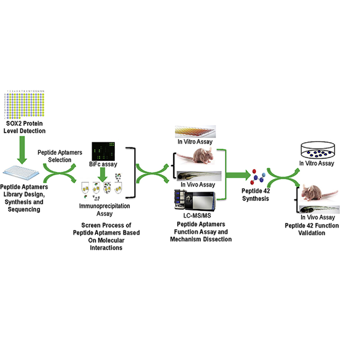
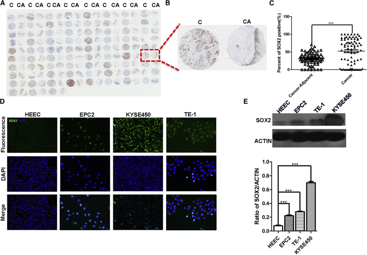
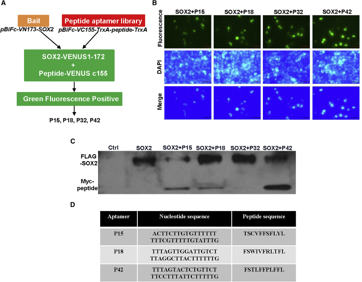
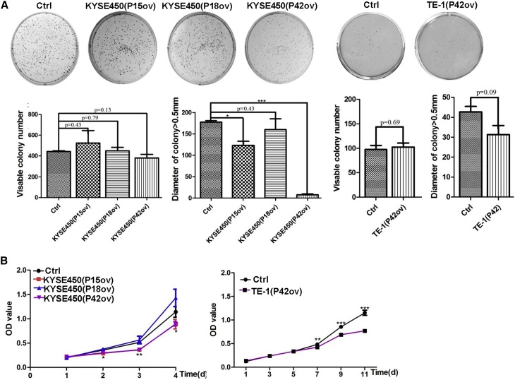
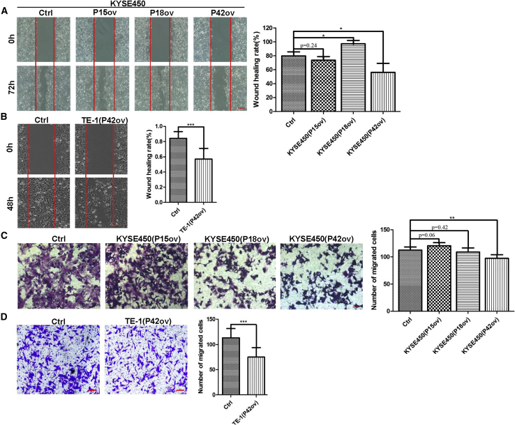
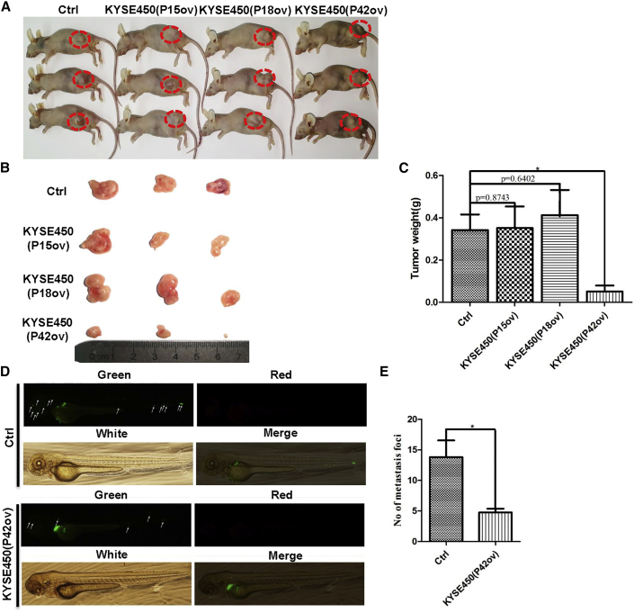
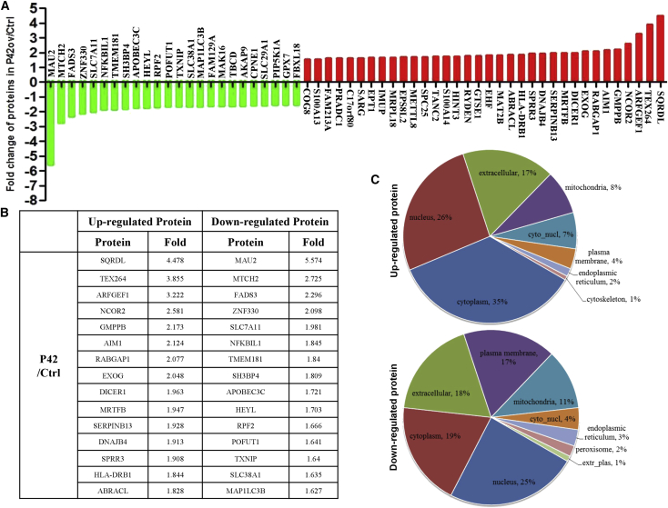
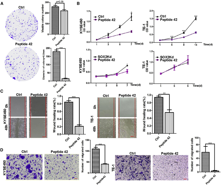
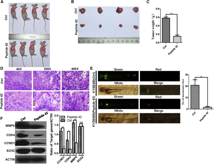
Esophageal squamous cell carcinoma (ESCC) is a predominant cancer type in developing countries such as China, where ESCC accounts for approximately 90% of esophageal malignancies. Lacking effective and targeted therapy contributes to the poor 5-year survival rate. Recent studies showed that about 30% of ESCC cases have high levels of SOX2. Herein, we aim to target this transcription factor with aptamer. We established a peptide aptamer library and then performed an unbiased screening to identify several peptide aptamers including P42 that can bind and inhibit SOX2 downstream target genes. We further found that P42 overexpression or incubation with a synthetic peptide 42 inhibited the proliferation, migration, and invasion of ESCC cells. Moreover, peptide 42 treatment inhibited the growth and metastasis of ESCC xenografts in mouse and zebrafish. Further analysis revealed that P42 overexpression led to alternations in the levels of proteins that are important for the proliferation and migration of ESCC cells. Taken together, our study identified the peptide 42 as a key inhibitor of SOX2 function, reducing the proliferation and migration of ESCC cells in vitro and in vivo, and thereby offering a potential therapy against ESCC.

Keywords: SOX2; drug screen; esophageal squamous cell carcinoma; peptide; peptide aptamers.


Figures

None
Graphical abstract
Figure 1
Figure 1
The Levels of SOX2 Are Increased in ESCC Samples and Cell Lines (A) Increased levels of SOX2 protein in ESCC samples (C) versus controls (CA) in tissue microarray. (B) A representative of ESCC/control pair. (C) The levels of SOX2 protein in ESCC cancer tissue are significantly higher than adjacent normal tissues (n = 75; p < 0.001). (D) SOX2 is enriched in ESCC cell lines (KYSE450, TE-1) and immortalized esophageal progenitor cell line (EPC2). Note SOX2 levels are relatively low in an esophageal epithelial cell line (HECC). Scale bars: 100 μm. (E) The levels of SOX2 protein were quantitated with western blot analysis. *p < 0.05, **p < 0.01, ***p < 0.001 versus control. Data are means ± SD.
Figure 2
Figure 2
Establishment of a Peptide Aptamer Library and Screening for Specific Peptides Aptamers that Target SOX2 Protein (A) Schematic of strategy generating peptide aptamer library. The vector expressing peptide aptamers was inserted, pBiFc-VC155-TrxA-MCS-TrxA, to establish the aptamer library, pBiFc-VC155-TrxA-peptide-TrxA, and SOX2-VENUS1-172 fusion protein is expressed with pBiFc-VN173-SOX2. If SOX2 binds potential aptamer, GFP will be produced based on BiFc and visualized with fluorescent microscope. (B) BiFc-based peptide aptamers P15, P18, P32, and P42 specifically bind SOX2 protein and generate green fluorescent signals in HEK293T cells. Scale bars: 50 μm. (C) Peptide aptamers P15, P18, and P42, but not P32, bind SOX2 protein as validated by co-immunoprecipitation. (D) The amino acid and nucleic acid sequences of peptide aptamers that specifically bind SOX2 protein.
Figure 3
Figure 3
P42 Aptamer Inhibits the Proliferation of ESCC Cells In Vitro (A) Ectopic expression of aptamers P42, P15, and P18 does not affect the colony numbers formed by KYSE450. However, the numbers of colonies with size larger than 0.5 mm are significantly reduced by aptamers P42 and P15, but not P18 (p < 0.05 for P15 aptamer and p < 0.001 for P42 aptamer; n = 3). Although it is statistically not significant, ectopic expression of P42 aptamer reduces the growth of colonies (size larger than 0.5 mm) formed by TE-1 cells (p > 0.05; n = 3). (B) Ectopic expression of aptamer P42 leads to reduced proliferation of KYSE450 and TE-1 cells as assessed by CCK8 assay (n = 3 per group; p < 0.05 for aptamers P15 and P42 in KYSE450 cells, p < 0.001 for aptamer P42 in TE-1 cells).
Figure 4
Figure 4
P42 Aptamer Inhibits the Migration and Invasion of ESCC Cells In Vitro (A and B) Ectopic expression of P42 aptamer inhibits the migration of (A) KYSE450 and (B) TE-1 cells as revealed by wound-healing assay (n = 3 per group; p < 0.05 for KYSE450 and p < 0.001 for TE-1 cells). Note P18 overexpression promotes the migration of KYSE450 cells. Scale bars: 500 μm. (C and D) P42 aptamer overexpression suppresses the invasion of (C) KYSE450 and (D) TE-1 cells in transwell assay (n = 3 per group; p < 0.01 for KYSE450 and p < 0.001 for TE-1 cells). Scale bars: 100 μm. *p < 0.05, **p < 0.01, ***p < 0.001 versus control. Data are means ± SD.
Figure 5
Figure 5
Ectopic Expression of P42 Aptamer Suppresses the Growth of Xenografts in Mice and Metastasis of KYSE450 Cells in Zebrafish (A and B) Representative tumors formed in mice following injection with KYSE450 cells that stably express P15, P18, or P42 aptamers. (C) Ectopic expression of P42 aptamer significantly reduces the weight of xenografts initiated by KYSE450 cells more than other peptide aptamers (n = 3 per group; p < 0.05). (D and E) P42 aptamer overexpression significantly inhibits the metastasis of KYSE450 cells more than control peptide aptamer along the body of zebrafish. Metastatic foci were examined with fluorescent microscopy (n = 5 per group; p < 0.05). *p < 0.05, **p < 0.01, ***p < 0.001 versus control. Data are means ± SD.
Figure 6
Figure 6
P42 Aptamer Overexpression Induces Changes in the Levels of Proteins that Have Been Implicated in Cancer Progression (A) Proteins that either increase or decrease by >1.5-fold upon ectopic expression of P42 aptamer in KYSE450 cells. (B) Top 15 proteins that are either upregulated or downregulated upon P42 aptamer ectopic expression. (C) Subcellular localization of the upregulated and downregulated proteins upon P42 aptamer ectopic expression.
Figure 7
Figure 7
Synthetic Peptide 42 (sP42) Inhibits the Proliferation, Migration, and Invasion of ESCC Cells In Vitro (A) sP42 application does not lead to reduction in the total number of KYSE450 colonies (p > 0.05), but the numbers of colonies larger than 0.5 mm are significantly reduced (n = 3 per group; p < 0.001). (B) Addition of sP42 to culture medium significantly reduces the proliferation of KYSE450 and TE-1 cells (n = 3 per group; p < 0.05 for KYSE450 cells and p < 0.001 for TE-1 cells). Note that the efficacy is comparable with SOX2 knockdown in KYSE450 and TE-1 cells (n = 3 per group; p < 0.05 for KYSE450 cells). (C) sP42 application leads to reduced migration of KYSE450 and TE-1 cells (n = 3 per group; p < 0.001 for KYSE450 cells and p < 0.01 for TE-1 cells). Scale bars: 500 μm. (D) sP42 application reduces the invasion of KYSE450 and TE-1 cells (n = 3 per group; p < 0.001 for KYSE450 cells and TE-1 cells). Scale bars: 100 μm. *p < 0.05, **p < 0.01, ***p < 0.001 versus control. Data are means ± SD.
Figure 8
Figure 8
sP42 Treatment Reduces the Growth of Xenografts Initiated by KYSE450 Cells and Inhibits the Metastasis of ESCC Cells in Zebrafish (A and B) sP42 treatment dramatically reduces the growth of xenografts than control peptide. sP42 was injected into xenografts every 2 days for 10 times. (C) sP42 treatment reduces the growth of KYSE450 cell-initiated xenograft more than control peptide (n = 4 per group; p < 0.01). (D) sP42 application promotes terminal differentiation of cancer cells with the increased presence of keratin pearls and intercellular bridges. Scale bars: 100 μm. (E) sP42 treatment significantly inhibits the metastasis of KYSE450 cells along the axis of zebrafish (n = 5 per group; p < 0.01). (F) sP42 treatment leads to reduced protein levels of CCND1, CDK4, and MMP8, but not SOX2 protein. *p < 0.05, **p < 0.01, ***p < 0.001 versus control. Data are means ± SD.

References

    1. Liang H., Fan J.H., Qiao Y.L. Epidemiology, etiology, and prevention of esophageal squamous cell carcinoma in China. Cancer Biol. Med. 2017;14:33–41. - PMC - PubMed
    1. Prabhu A., Obi K.O., Rubenstein J.H. The synergistic effects of alcohol and tobacco consumption on the risk of esophageal squamous cell carcinoma: a meta-analysis. Am. J. Gastroenterol. 2014;109:822–827. - PubMed
    1. Liu X., Wang X., Lin S., Yuan J., Yu I.T. Dietary patterns and oesophageal squamous cell carcinoma: a systematic review and meta-analysis. Br. J. Cancer. 2014;110:2785–2795. - PMC - PubMed
    1. Chang F., Syrjänen S., Wang L., Syrjänen K. Infectious agents in the etiology of esophageal cancer. Gastroenterology. 1992;103:1336–1348. - PubMed
    1. Chen T., Cheng H., Chen X., Yuan Z., Yang X., Zhuang M., Lu M., Jin L., Ye W. Family history of esophageal cancer increases the risk of esophageal squamous cell carcinoma. Sci. Rep. 2015;5:16038. - PMC - PubMed

Publication types

MeSH terms

LinkOut - more resources