Review
. 2020 May 1;1862(5):183206.
doi: 10.1016/j.bbamem.2020.183206. Epub 2020 Jan 25.

Chapter 22: Structural and signaling functions of integrins

Affiliations
Review

Chapter 22: Structural and signaling functions of integrins

Yasmin A Kadry et al. Biochim Biophys Acta Biomembr. .

Abstract

The integrin family of transmembrane adhesion receptors is essential for sensing and adhering to the extracellular environment. Integrins are heterodimers composed of non-covalently associated α and β subunits that engage extracellular matrix proteins and couple to intracellular signaling and cytoskeletal complexes. Humans have 24 different integrin heterodimers with differing ligand binding specificities and non-redundant functions. Complex structural rearrangements control the ability of integrins to engage ligands and to activate diverse downstream signaling networks, modulating cell adhesion and dynamics, processes which are crucial for metazoan life and development. Here we review the structural and signaling functions of integrins focusing on recent advances which have enhanced our understanding of how integrins are activated and regulated, and the cytoplasmic signaling networks downstream of integrins.

Keywords: Cytoskeleton; Focal adhesion; Integrin; Kindlin; Talin.


Conflict of interest statement

Declaration of competing interest The authors declare that they have no known competing financial interests or personal relationships that could have appeared to influence the work reported in this paper.

Figures
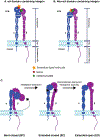
Figure 1:
Figure 1:. Integrin domain architecture and conformational states.
(A,B) Schematic of the domain architecture of an αA domain-containing (A) and non-αA domain containing (B) integrin heterodimer in the active (extended-open) state. Metal-ion binding sites (MIDAS, ADMIDAS, and SyMBS) are indicated. (C) Cartoon depiction of the conformational transitions of the integrin heterodimer from a bent-closed (BC) to an extended-closed (EC) and finally to an extended-open (EO) conformation.
Figure 2:
Figure 2:. The integrin
α-TMD and β-TMD. Solution NMR structure of the α-TMD and β-TMD of αIIbβ3 (PDB ID: 2K9J), with the outer membrane clasp (OMC) and inner membrane clasp (IMC) as indicated [41].
Figure 3:
Figure 3:. Sequence alignment of β-tail cytoplasmic domains.
Sequence alignment of human β-integrin cytoplasmic tail protein sequences. Conserved ‘NP(I/L)(Y/F)’, ‘Nxx(Y/F)’, and Serine/Threonine rich motifs are indicated. Alignment was generated using the ESPript web server, and ESPript-calculated conserved residues are colored in red and boxed in blue [61]. β4 was omitted from this alignment due to its unusually large length, and β8 was excluded due to a lack of strong conservation with the other β subunits in the alignment.
Figure 4:
Figure 4:. The integrin activation process.
A partial overview of the integrin activation process. First, intracellular adaptor recruitment drives conformational activation and the transition from the bent-closed to extended-open state. Following this, integrin clustering and focal adhesion assembly begins. Finally, focal adhesion disassembly/integrin inactivation is promoted by the recruitment of endocytic/inhibitory adaptors to the cytoplasmic tails.
Figure 5:
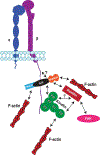
Figure 5:. The kindlin-IPP-paxillin interactome.
A summary of the major interactome connections between kindlin, paxillin and the IPP complex identified to date. Kindlin has been reported to interact with the IPP complex [146,150,161], F-actin [151], and paxillin at two distinct sites in the F0 [148,149,186,188] and PH domains [148,188], although only the kindlin F0-paxillin LIM4 interaction has been verified structurally [186]. Meanwhile, interactions between the IPP complex (through parvin) and paxillin [172,173], as well as between the IPP complex and F-actin [166], have also been reported, creating a complex signaling network centered around kindlins. Moreover, the extent of the signaling network is enhanced by reported interactions between paxillin and focal adhesion kinase [183]. Double-headed arrows indicate a reported interaction between the two binding partners.
Figure 6:
Figure 6:. Hemidesmosomes vs. focal adhesions.
(A) Hemidesmosomes connect integrins to the intermediate filament cytoskeleton, while (B) focal adhesions link integrins to the F-actin cytoskeleton, both directly and indirectly.

References

    1. Hegde S, Raghavan S, A skin-depth analysis of integrins: Role of the integrin network in health and disease, Cell Commun. Adhes 20 (2013) 155–169. doi:10.3109/15419061.2013.854334. - DOI - PubMed
    1. Winograd-Katz SE, Fässler R, Geiger B, Legate KR, The integrin adhesome: From genes and proteins to human disease, Nat. Rev. Mol. Cell Biol 15 (2014) 273–288. doi:10.1038/nrm3769. - DOI - PubMed
    1. Plow EF, Meller J, Byzova TV, Integrin function in vascular biology: A view from 2013, Curr. Opin. Hematol 21 (2014) 241–247. doi:10.1097/MOH.0000000000000042. - DOI - PMC - PubMed
    1. Wickström SA, Radovanac K, Fässler R, Genetic analyses of integrin signaling, Cold Spring Harb. Perspect. Biol 3 (2011) 1–22. doi:10.1101/cshperspect.a005116. - DOI - PMC - PubMed
    1. Goodman SL, Picard M, Integrins as therapeutic targets, Trends Pharmacol. Sci 33 (2012) 405–412. doi:10.1016/j.tips.2012.04.002. - DOI - PubMed

Publication types

LinkOut - more resources