

Systemic distribution of progesterone receptor subtypes in human tissues
- PMID: 31991170
- PMCID: PMC9968951
- DOI: 10.1016/j.jsbmb.2020.105599
Systemic distribution of progesterone receptor subtypes in human tissues
Abstract
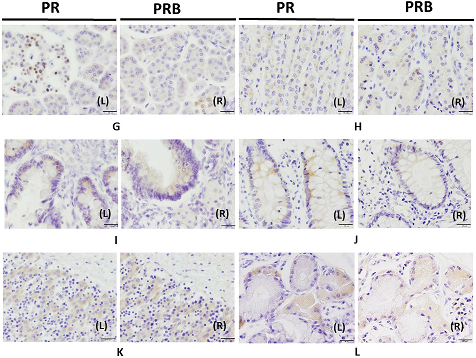
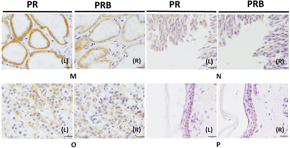
Progesterone receptor (PR) is expressed in a wide variety of human tissues, including both reproductive and non-reproductive tissues. Upon binding to the PR, progesterone can display several non-reproductive functions, including neurosteroid activity in the central nervous system, inhibition of smooth muscle contractile activity in the gastrointestinal tract, and regulating the development and maturation of the lung. PR exists as two major isoforms, PRA and PRB. Differential expression of these PR isoforms reportedly contributes to different biological activities of the hormone. However, the distribution of the PR isoforms in human tissues has remained virtually unexplored. In this study, we immunolocalized PR expression in various human tissues using PR (1294) specific antibody, which is capable of detecting both PRA and PRB, and PRB (250H11) specific antibody. Tissues from the uterus, ovary, breast, placenta, prostate, testis, cerebrum, cerebellum, pituitary, spinal cord, esophagus, stomach, small intestine, colon, pancreas, liver, kidney, urinary bladder, lung, heart, aorta, thymus, adrenal gland, thyroid, spleen, skin, and bone were examined in four different age groups (fetal, pediatric, young, and old) in male and female subjects. PR and PRB were detected in the nuclei of cells in the female reproductive system, in both the nuclei and cytoplasm of pituitary gland and pancreatic acinar cells, and only in the cytoplasm of cells in the testis, stomach, small intestine, colon, liver, kidney, urinary bladder, lung, adrenal gland, and skin. Of particular interest, total PRB expression overlapped with that of total PR expression in most tissues but was negative in the female fetal reproductive system. The findings indicate that progesterone could affect diverse human organs differently than from reproductive organs. These findings provide new insights into the novel biological roles of progesterone in non-reproductive organs.
Keywords: Human tissues; Progesterone receptor; Progesterone receptor isoform B; Systemic distribution.
Copyright © 2020 Elsevier Ltd. All rights reserved.
Conflict of interest statement
Declaration of Competing Interest Authors report no conflict of interest.
Figures

References
-
- Ferenczy A, Bertrand G, Gelfand MM. Proliferation kinetics of human endometrium during the normal menstrual cycle. American journal of obstetrics and gynecology. 1979;133(8):859–67. - PubMed
-
- Michalas S, Loutradis D, Drakakis P, Kallianidis K, Milingos S, Deligeoroglou E, et al. A flexible protocol for the induction of recipient endometrial cycles in an oocyte donation programme. Human reproduction (Oxford, England). 1996;11(5):1063–6. - PubMed
-
- Nosarka S, Kruger T, Siebert I, Grove D. Luteal phase support in in vitro fertilization: meta-analysis of randomized trials. Gynecologic and obstetric investigation. 2005;60(2):67–74. https://doi.org: 10.1159/000084546. - DOI - PubMed
-
- Navot D, Anderson TL, Droesch K, Scott RT, Kreiner D, Rosenwaks Z. Hormonal manipulation of endometrial maturation. The Journal of clinical endocrinology and metabolism. 1989;68(4):801–7. https://doi.org: 10.1210/jcem-68-4-801. - DOI - PubMed
-
- Park O-K, Mayo KE. Transient expression of progesterone receptor messenger RNA in ovarian granuiosa cells after the preovulatory luteinizing hormone surge. Molecular endocrinology. 1991;5(7):967–78. - PubMed
Publication types
MeSH terms
Substances
Grants and funding
LinkOut - more resources
Full Text Sources
Research Materials
