

White Matter Brain Development after Exposure to Circulating Cell-Free Hemoglobin and Hyperoxia in a Rat Pup Model
- PMID: 31991415
- PMCID: PMC7212701
- DOI: 10.1159/000505206
White Matter Brain Development after Exposure to Circulating Cell-Free Hemoglobin and Hyperoxia in a Rat Pup Model
Abstract
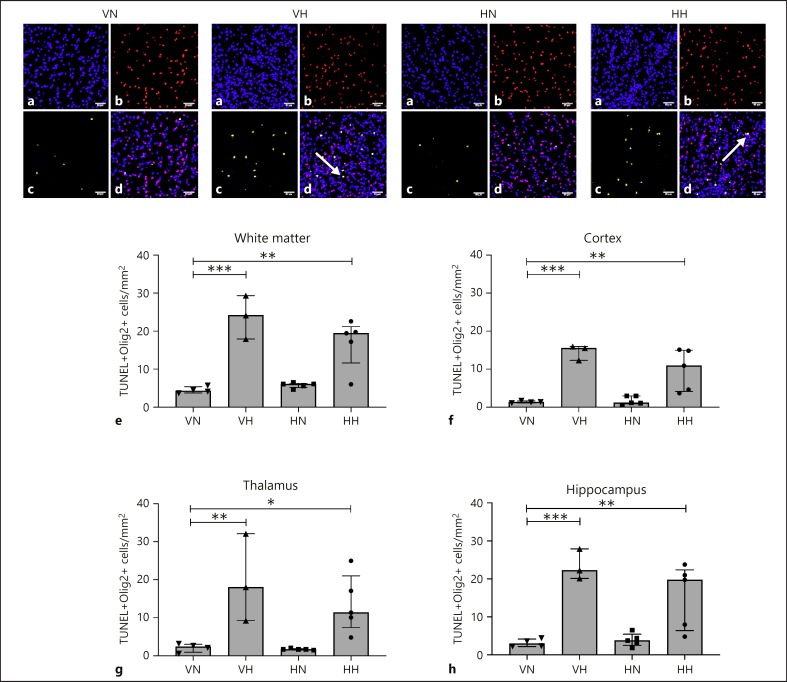
Neonates born with critical congenital heart defects are at risk of diffuse white matter injuries and neurodevelopmental impairments. This study aimed to determine the impact of circulating cell-free hemoglobin and hyperoxia, both present during cardiopulmonary bypass circulation, on white matter brain development. Postnatal day 6 rat pups were injected intraperitoneally with cell-free Hb or vehicle and exposed to hyperoxia (fiO2 = 0.8) or normoxia (fiO2 = 0.21) for 24 h. We evaluated apoptosis, myelination, and oligodendrocyte maturation with immunohistochemistry, gene and protein analyses, and in vivo diffusion tensor magnetic resonance imaging (MRI). Consistent with previous studies, we found an increase in apoptosis of oligodendrocytes as determined by TUNEL+ staining in Olig2+ cells in white matter, cortex, thalamus, and hippocampus following exposure to hyperoxia with no additional effect of cell-free Hb. A transient increase in the mRNA expression of intercellular adhesion molecule 1 at 6 h was observed following combined exposure to cell-free Hb and hyperoxia. No indications of oligodendrocyte maturational delay or hypomyelination were observed after either insult, delivered separately or combined, as determined by immunohistochemistry, Western blot, and diffusion tensor MRI. In our model, exposure to circulatory cell-free Hb, with or without concomitant hyperoxia, did not significantly alter brain white matter development.
Keywords: Cardiopulmonary bypass; Cell-free hemoglobin; Hyperoxia; Oligodendrocyte; White matter.
© 2020 S. Karger AG, Basel.
Conflict of interest statement
The authors declare no conflicts of interest.
Figures


Similar articles
-
Cardiopulmonary bypass in the newborn: effects of circulatory cell-free hemoglobin and hyperoxia evaluated in a novel rat pup model.Intensive Care Med Exp. 2017 Oct 4;5(1):45. doi: 10.1186/s40635-017-0153-2. Intensive Care Med Exp. 2017. PMID: 28980221 Free PMC article.
-
Fingolimod protects against neonatal white matter damage and long-term cognitive deficits caused by hyperoxia.Brain Behav Immun. 2016 Feb;52:106-119. doi: 10.1016/j.bbi.2015.10.004. Epub 2015 Oct 21. Brain Behav Immun. 2016. PMID: 26456693
-
Erythropoietin Restores Long-Term Neurocognitive Function Involving Mechanisms of Neuronal Plasticity in a Model of Hyperoxia-Induced Preterm Brain Injury.Oxid Med Cell Longev. 2016;2016:9247493. doi: 10.1155/2016/9247493. Epub 2016 Jul 14. Oxid Med Cell Longev. 2016. PMID: 27493706 Free PMC article.
-
Hyperoxia and the Immature Brain.Dev Neurosci. 2016;38(5):311-330. doi: 10.1159/000454917. Epub 2017 Feb 3. Dev Neurosci. 2016. PMID: 28152539 Review.
-
Diffusion tensor imaging for understanding brain development in early life.Annu Rev Psychol. 2015 Jan 3;66:853-76. doi: 10.1146/annurev-psych-010814-015340. Annu Rev Psychol. 2015. PMID: 25559117 Free PMC article. Review.
Cited by
-
Dynamic Changes of Endogenic or Exogenic β-Carboline Alkaloid Harmine in Different Mammals and Human in vivo at Developmental and Physiological States.Front Aging Neurosci. 2022 Jan 14;13:773638. doi: 10.3389/fnagi.2021.773638. eCollection 2021. Front Aging Neurosci. 2022. PMID: 35095466 Free PMC article.
-
Perinatal Hyperoxia and Developmental Consequences on the Lung-Brain Axis.Oxid Med Cell Longev. 2022 Feb 24;2022:5784146. doi: 10.1155/2022/5784146. eCollection 2022. Oxid Med Cell Longev. 2022. PMID: 35251477 Free PMC article. Review.
-
Cerebral Myelination in a Bronchopulmonary Dysplasia Murine Model.Children (Basel). 2023 Jul 31;10(8):1321. doi: 10.3390/children10081321. Children (Basel). 2023. PMID: 37628321 Free PMC article.
-
Mechanistic advances of hyperoxia-induced immature brain injury.Heliyon. 2024 Apr 22;10(9):e30005. doi: 10.1016/j.heliyon.2024.e30005. eCollection 2024 May 15. Heliyon. 2024. PMID: 38694048 Free PMC article. Review.
References
-
- Beca J, Gunn JK, Coleman L, Hope A, Reed PW, Hunt RW, et al. New white matter brain injury after infant heart surgery is associated with diagnostic group and the use of circulatory arrest. Circulation. 2013 Mar;127((9)):971–9. - PubMed
-
- Ricci Z, Pezzella C, Romagnoli S, Iodice F, Haiberger R, Carotti A, et al. High levels of free haemoglobin in neonates and infants undergoing surgery on cardiopulmonary bypass. Interact Cardiovasc Thorac Surg. 2014 Aug;19((2)):183–7. - PubMed
Publication types
MeSH terms
Substances
LinkOut - more resources
Full Text Sources
Other Literature Sources
