Review
. 2020 Feb 29;43(2):126-138.
doi: 10.14348/molcells.2019.0301.

RUNX1 Dosage in Development and Cancer

Affiliations
Review

RUNX1 Dosage in Development and Cancer

Michael Lie-A-Ling et al. Mol Cells. .

Abstract

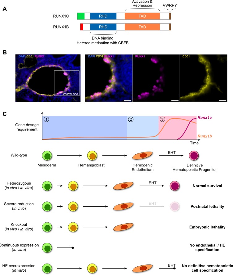
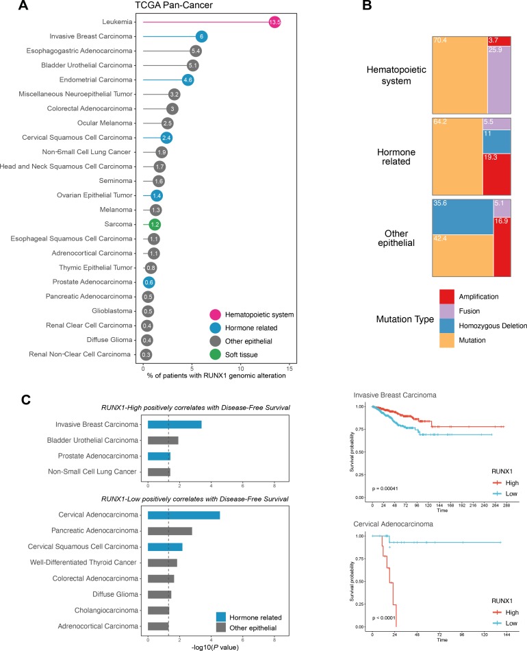
The transcription factor RUNX1 first came to prominence due to its involvement in the t(8;21) translocation in acute myeloid leukemia (AML). Since this discovery, RUNX1 has been shown to play important roles not only in leukemia but also in the ontogeny of the normal hematopoietic system. Although it is currently still challenging to fully assess the different parameters regulating RUNX1 dosage, it has become clear that the dose of RUNX1 can greatly affect both leukemia and normal hematopoietic development. It is also becoming evident that varying levels of RUNX1 expression can be used as markers of tumor progression not only in the hematopoietic system, but also in non-hematopoietic cancers. Here, we provide an overview of the current knowledge of the effects of RUNX1 dosage in normal development of both hematopoietic and epithelial tissues and their associated cancers.

Keywords: development; dosage; hematopoiesis; runx1; tumorigenesis.


Conflict of interest statement

Disclosure

The authors have no potential conflicts of interest to disclose.

Figures

Fig. 1
Fig. 1. RUNX1 dosage in hematopoietic development.
(A) Schematic representation of the two most abundant RUNX1 isoforms. Except for the most N-terminal sequence (RUNX1C N-terminal in green, RUNX1B N-terminal in red) the proteins are identical and they both contain the highly conserved Runt homology domain (RHD, blue) followed by a transactivation domain (TAD, orange) which is flanked by inhibitory regions. The C-terminal inhibitory region contains a highly conserved VWRPY motif (brown). (B) Immunofluorescence on the AGM of a E10.5 mouse embryo. The dorsal aortic endothelial cells are marked by the endothelial marker CD31 (yellow). The majority of the cells on the ventral side of the dorsal aorta (constituting both endothelial and rare HE cells) are positive for the RUNX1 protein (magenta). Scale bars = 20 μm. (C) Current model of RUNX1 dosage in hematopoietic development. Top: RUNX1 dosage requirement can be divided in three phases. Phase ①: early in differentiation RUNX1 is not required but its (low) dose influences the timing and dynamics of HE cells appearance. Phase ②: although RUNX1 levels are still low in HE cells, its presence is required for the initiation of the EHT. Phase ③: an increased dose of RUNX1 is required for the completion of EHT and the generation of the first mature hematopoietic cells. The whole differentiation process is predominantly controlled by the RUNX1b isoform. Bottom: schematic overview of the currently available phenotypic data on RUNX1 dosage during the establishment of the hematopoietic system in the embryo.
Fig. 2
Fig. 2. Meta-analysis of RUNX1 alterations and prognostic value in the TCGA PanCancer atlas.
(A) Frequency of RUNX1 genomic alterations across the TCGA PanCancer atlas. Cancers with no alterations were excluded. Cancers affecting the hematopoietic system are colored in pink, hormone related cancers in blue, cancers of soft tissues in green, and other epithelial cancers in grey. (B) Proportion of RUNX1 amplification, homozygous deletion, fusion and mutation in cancers affecting the hematopoietic system, hormone related cancers, and additional epithelial cancers. Soft tissue cancers were excluded from these analyses due to the small number of patients affected. (C) Prognostic value of RUNX1 mRNA expression using the TCGA PanCancer Atlas expression data, in terms of Disease-Free Survival. Datasets of the TCGA PanCancer Atlas were downloaded from cBioPortal (https://www.cbioportal.org/). Briefly, patients were split in RUNX1-High and RUNX1-Low groups using the “surv_cutpoint” function of the “survminer” R package (“minprop” argument set to 0.1). Cancers were then separated into two groups, depending on whether RUNX1-High and RUNX1-Low groups are significantly associated with a better prognosis (P value < 0.05 using the univariate log-rank test). Representative examples of the corresponding Kaplan–Meier curves are shown for the Invasive Breast Carcinoma and Cervical Adenocarcinoma datasets (defined by the “Cancer Type” column of the TCGA PanCancer Atlas clinical data).

References

    1. Aikawa Y., Nguyen L.A., Isono K., Takakura N., Tagata Y., Schmitz M.L., Koseki H., Kitabayashi I. Roles of HIPK1 and HIPK2 in AML1- and p300-dependent transcription, hematopoiesis and blood vessel formation. EMBO J. 2006;25:3955–3965. doi: 10.1038/sj.emboj.7601273. - DOI - PMC - PubMed
    1. Angelos M.G., Abrahante J.E., Blum R.H., Kaufman D.S. Single cell resolution of human hematoendothelial cells defines transcriptional signatures of hemogenic endothelium. Stem Cells. 2018;36:206–217. doi: 10.1002/stem.2739. - DOI - PMC - PubMed
    1. Aronson B.D., Fisher A.L., Blechman K., Caudy M., Gergen J.P. Groucho-dependent and -independent repression activities of Runt domain proteins. Mol. Cell. Biol. 1997;17:5581–5587. doi: 10.1128/MCB.17.9.5581. - DOI - PMC - PubMed
    1. Bae S.C., Yamaguchi-Iwai Y., Ogawa E., Maruyama M., Inuzuka M., Kagoshima H., Shigesada K., Satake M., Ito Y. Isolation of PEBP2 alpha B cDNA representing the mouse homolog of human acute myeloid leukemia gene, AML1. Oncogene. 1993;8:809–81. - PubMed
    1. Banach-Petrosky W., Jessen W.J., Ouyang X., Gao H., Rao J., Quinn J., Aronow B.J., Abate-Shen C. Prolonged exposure to reduced levels of androgen accelerates prostate cancer progression in Nkx3.1; Pten mutant mice. Cancer Res. 2007;67:9089–9096. doi: 10.1158/0008-5472.CAN-07-2887. - DOI - PubMed

MeSH terms

Substances