Review
. 2020 Jan 24;10(2):179.
doi: 10.3390/biom10020179.

FAK Structure and Regulation by Membrane Interactions and Force in Focal Adhesions

Affiliations
Review

FAK Structure and Regulation by Membrane Interactions and Force in Focal Adhesions

Paula Tapial Martínez et al. Biomolecules. .

Abstract

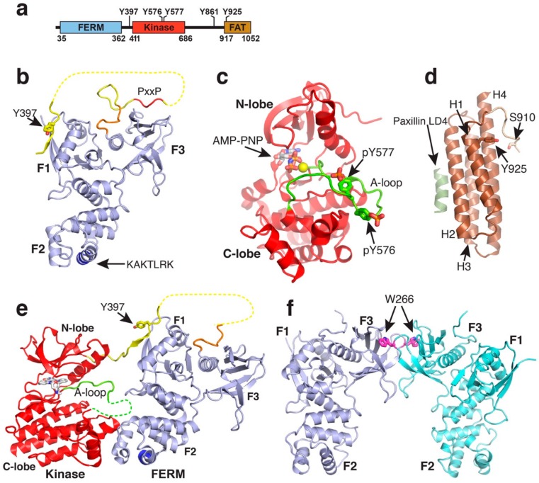
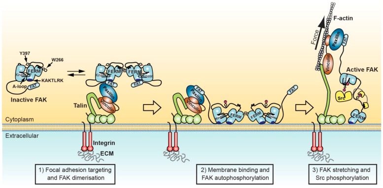
Focal adhesion kinase (FAK) is a non-receptor tyrosine kinase with key roles in the regulation of cell adhesion migration, proliferation and survival. In cancer FAK is a major driver of invasion and metastasis and its upregulation is associated with poor patient prognosis. FAK is autoinhibited in the cytosol, but activated upon localisation into a protein complex, known as focal adhesion complex. This complex forms upon cell adhesion to the extracellular matrix (ECM) at the cytoplasmic side of the plasma membrane at sites of ECM attachment. FAK is anchored to the complex via multiple sites, including direct interactions with specific membrane lipids and connector proteins that attach focal adhesions to the actin cytoskeleton. In migrating cells, the contraction of actomyosin stress fibres attached to the focal adhesion complex apply a force to the complex, which is likely transmitted to the FAK protein, causing stretching of the FAK molecule. In this review we discuss the current knowledge of the FAK structure and how specific structural features are involved in the regulation of FAK signalling. We focus on two major regulatory mechanisms known to contribute to FAK activation, namely interactions with membrane lipids and stretching forces applied to FAK, and discuss how they might induce structural changes that facilitate FAK activation.

Keywords: cell adhesion; cell signalling; focal adhesion kinase; mechanobiology; membrane interactions; structural biology.


Conflict of interest statement

The authors declare no conflict of interest.

Figures

Figure 1
Figure 1
Structures of FAK domains. (a) Schematic domain structure of FAK. Domain boundaries and regulatory phosphorylation sites are indicated. (b) Crystal structure of the FAK FERM domain containing the F1, F1 and F3 lobes (PDB accession 2AL6). The 10 C-terminal residues integral to the FAK FERM domain, but not in ERM FERM domains, are coloured in orange. The linker is in yellow with the PxxP motif known to interact with the Src SH3 domain in red. The Y397 autophosphorylation site in the linker and the KAKTLRK motif in the F2 lobe (blue) are labelled. (c) Structure of the active FAK kinase domain with Y576 and Y577 in the activation loop (A-loop) phosphorylated (PDB accession 2J0L). The A-loop is coloured in green. A non-hydrolysable ATP analogue (AMP-PNP) and a Mg2+ ion (yellow sphere) are bound to the active site. (d) Structure of the FAT domain bound to the paxillin LD4 peptide (PDB accession 1OW7). The paxillin LD4 peptide is in olive green and the N-terminal FAT extension in tan. Helices H1-H4 and the S910 and Y925 phosphorylation sites are labelled. (e) Crystal structure of the FERM-kinase region of FAK in the autoinhibited conformation (PDB accession 2J0J). Colouring is as in panels (a,b). (f) FERM dimer mediated by W266 interactions in the F3 lobe as observed in various crystal structures (shown from PDB accession 2AEH).
Figure 2
Figure 2
Model for FAK activation in focal adhesions mediated by membrane interactions and force. Left: FAK can form dimers via F3 lobe interactions involving W266. FAK dimer formation is likely promoted upon recruitment into focal adhesions by increased local concentration and potentially by paxillin-FAT interactions that synergise with FAT-FERM interactions to stabilise FAK dimers. Middle: FAK interacts with nearby PIP2 rich membranes via the KAKTLRK basic patch in the FERM F2 lobe. Increased avidity for PIP2 of FAK dimers might enhance membrane attachment. Membrane interactions induce conformational changes that allow simultaneous interactions of FERM and kinase domains to the membrane and expose the FERM-kinase linker for efficient autophosphorylation on Y397. Right: Src binds with its SH2 domain to the phosphorylated Y397 site and with the SH3 domain to the PxxP motif in the FERM-kinase linker. Stretching forces applied to FAK by contracting actomyosin fibres cause release of the FAK kinase domain from FERM and membrane interactions, exposing the activation loop for efficient phosphorylation by Src, resulting in FAK activation.

References

    1. Hanks S.K., Calalb M.B., Harper M.C., Patel S.K. Focal adhesion protein-tyrosine kinase phosphorylated in response to cell attachment to fibronectin. Proc. Natl. Acad. Sci. USA. 1992;89:8487–8491. doi: 10.1073/pnas.89.18.8487. - DOI - PMC - PubMed
    1. Schaller M.D., Borgman C.A., Cobb B.S., Vines R.R., Reynolds A.B., Parsons J.T. pp125FAK a structurally distinctive protein-tyrosine kinase associated with focal adhesions. Proc. Natl. Acad. Sci. USA. 1992;89:5192–5196. doi: 10.1073/pnas.89.11.5192. - DOI - PMC - PubMed
    1. Ilic D., Furuta Y., Kanazawa S., Talkeda N., Sobue K., Nakatsuji N., Nomura S., Fujimoto J., Okada M., Yamamoto T. Reduced cell motility and enhanced focal adhesion contact formation in cells from FAK-deficient mice. Nature. 1995;377:539–544. - PubMed
    1. Lim S.T., Chen X.L., Tomar A., Miller N.L., Yoo J., Schlaepfer D.D. Knock-in mutation reveals an essential role for focal adhesion kinase activity in blood vessel morphogenesis and cell motility-polarity but not cell proliferation. J. Biol. Chem. 2010;285:21526–21536. doi: 10.1074/jbc.M110.129999. - DOI - PMC - PubMed
    1. Matsuura S., Kondo S., Suga K., Kinoshita Y., Urushihara M., Kagami S. Expression of focal adhesion proteins in the developing rat kidney. J. Histochem. Cytochem. 2011;59:864–874. doi: 10.1369/0022155411413929. - DOI - PMC - PubMed

MeSH terms

Substances