. 2020 Jan 24;21(3):763.
doi: 10.3390/ijms21030763.

Advanced Methodology and Preliminary Measurements of Molecular and Mechanical Properties of Heart Valves under Dynamic Strain

Affiliations

Advanced Methodology and Preliminary Measurements of Molecular and Mechanical Properties of Heart Valves under Dynamic Strain

Rama S Madhurapantula et al. Int J Mol Sci. .

Abstract

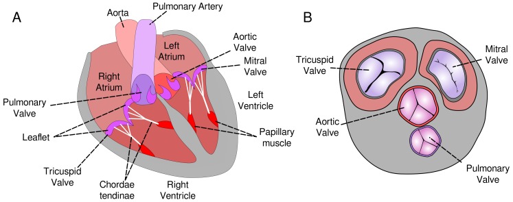
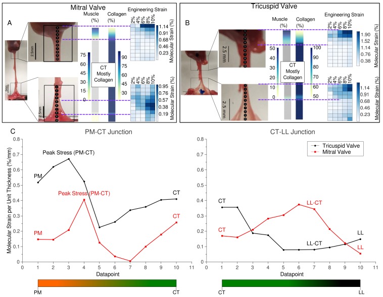
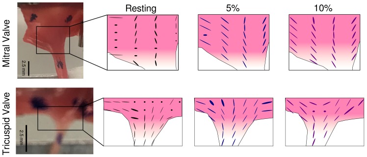
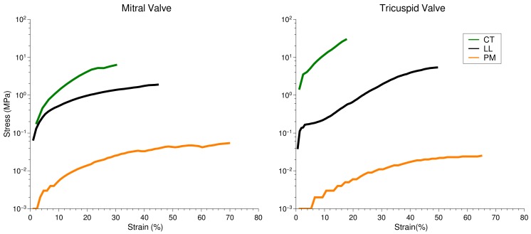
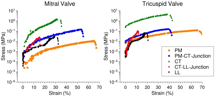
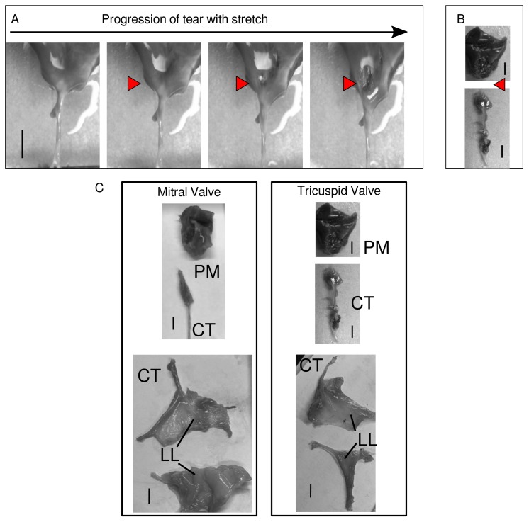
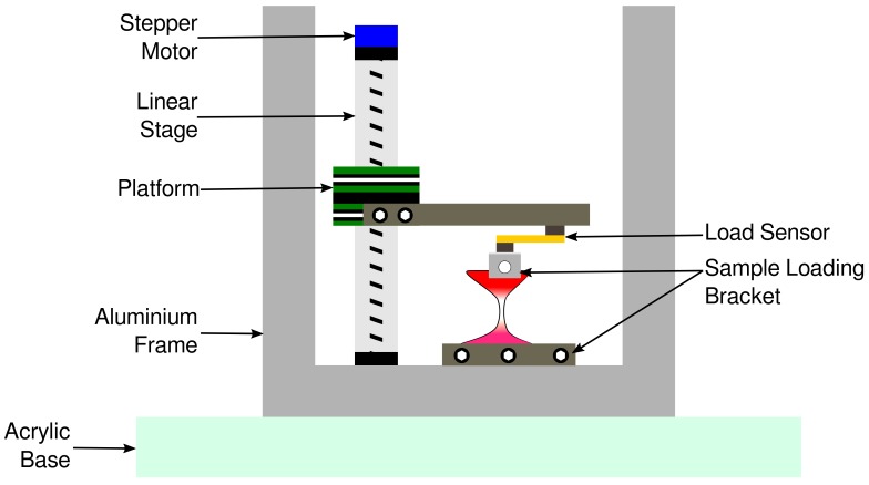
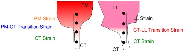
Mammalian heart valves are soft tissue assemblies with multi-scale material properties. This is because they are constructs comprising both muscle and non-contractile extracellular matrix proteins (such as collagens and proteoglycans) and transition regions where one form of tissue structure becomes another, significantly different form. The leaflets of the mitral and tricuspid valves are connected to chordae tendinae which, in turn, bind through papillary muscles to the cardiac wall of the ventricle. The transition regions between these tissue subsets are complex and diffuse. Their material composition and mechanical properties have not been previously described with both micro and nanoscopic data recorded simultaneously, as reported here. Annotating the mechanical characteristics of these tissue transitions will be of great value in developing novel implants, improving the state of the surgical simulators and advancing robot-assisted surgery. We present here developments in multi-scale methodology that produce data that can relate mechanical properties to molecular structure using scanning X-ray diffraction. We correlate these data to corresponding tissue level (macro and microscopic) stress and strain, with particular emphasis on the transition regions and present analyses to indicate points of possible failure in these tissues.

Keywords: X-ray diffraction scanning; heart valves; stress–strain relations; tissue transition regions; valve failure.


Conflict of interest statement

The authors disclose a perceived conflict of interest for this edition in that the senior author (J.P.R.O.O) is an editor of the “Molecular Tissue Responses to Mechanical Loading” special edition and a member of the editorial board. The co-editor, Ashley Eidsmore, was also a co-author of a manuscript with the lead and senior authors within the last 12 months. The authors declare no such relationship or affiliation with any of the suggested reviewers, nor any other known conflicts or perceived conflicts.

Figures

Figure 1
Figure 1
Illustration of the human heart valves. The leaflet of the MV and TV is held in position by CT that extend into the PM, which attach on to the wall of the heart. (A) Lateral (frontal) cross-section, (B) Transverse cross-section.
Figure 2
Figure 2
Tissue composition and molecular strain vs. applied engineering strain maps from ‘bisected’ MV and TV samples. (A,B) Composition maps showing muscle and collagen percentages along the PM–CT and CT–LL junctions. These transitions are diffuse in nature. Particularly in the PM–CT junction, the CT extends into the PM by a few millimeters. There is a near-linear decrease in collagen percentage, going further into the PM, starting at the beginning of the PM. These transition regions are marked by the purple dotted lines. A comparison of engineering strain (applied stretch/strain) vs. the molecular strain (calculated from changes in collagen D-period elongation) is shown as heatmaps for both the transitions from MV and TV. An increase in molecular strain is observed, particularly in the PM–CT junction. Similar localization of molecular strain is observed in the LL–CT transition of the MV but not the TV. (C) Tracking of molecular strain per unit thickness along the PM–CT and the CT–LL transition regions of MV and TV. The molecular strain data are obtained from the 10% engineering strain series in each case. This measure provides with a representation of local strain distribution, which may be helpful to determine the point of breakage. As observed, the PM–CT junction assumes highest molecular strain (peak stress labeled on the plot). A second potential point of failure is observed in the MV, at the LL–CT transition. However, the overall molecular strain per unit length experienced by the LL–CT junction is lower than that of the PM–CT.
Figure 3
Figure 3
Local collagen fiber alignment calculated from 2-D XRD scans on ‘bisected’ CT–LL samples from TV and LV. The alignment at each datapoint is represented as an ellipse. As is observed with the 5% and 10% stretch series, more fibers become more aligned with increased overall strain applied on the sample.
Figure 4
Figure 4
Stress vs. strain evaluation of individual sample elements from MV and TV samples tri-sected so as to measure pure tissue sub-types independently.
Figure 5
Figure 5
Stress vs. strain evaluation of individual sample segments from MV and TV bisected samples so as to measure these properties in the transition regions in addition to that of pure regions connected to the transition. Stress vs. strain plots were calculated by tracking the movement of fiducial marker. The PM is the most compliant element in the assembly, assuming most strain over the least amount of stress. The CT is the most tensile element, assuming least strain for most stress. The PM–CT junction, in both valves, has the second highest stress for strain. Several points were recorded after the beginning point of sample failure showing the steep decline in the plot of strain vs. stress for each component. These data were used to help determine the ultimate tensile strength (stress) of each tissue component.
Figure 6
Figure 6
(A) Tear in the PM–CT junction of the TV. The red arrowheads show the beginning and progression of the tear in the sample with stretch. (B) shows a picture of a torn sample and the point of detachment. (C) Post-tear. Note in (B) and (C), the PM–CT sample from TV were recorded in one image that was modified to place the PM and CT elements closer together for direct comparison to MV image. The sample itself was not modified in the image. Scale bar in all panels shows 5 mm.
Figure 7
Figure 7
Sample strain apparatus. The stepper motor actuates the stage up to strain the samples loaded between the sample loading brackets. The aluminum frame is attached to a 1/8th inch clear acrylic sheet which acts as a door (not shown in the illustration) to load samples. The thick acrylic base is used to stabilize the rig during data acquisition and also for mounting the rig at the X-ray beamline on an XY sample positioner.
Figure 8
Figure 8
An illustration of location of fiducial markers placed on samples for tracking using the TrackMate plugin on Image J. Markers are placed so that each region (PM, CT and LL) have at least have one marker and one on each side of the visible transition in each sample type.

References

    1. Nkomo V.T., Gardin J.M., Skelton T.N., Gottdiener J.S., Scott C.G., Enriquez-Sarano M. Burden of valvular heart diseases: A population-based study. Lancet. 2006;368:1005–1011. doi: 10.1016/S0140-6736(06)69208-8. - DOI - PubMed
    1. Vukanovic-Criley J.M., Criley S., Warde C.M., Boker J.R., Guevara-Matheus L., Churchill W.H., Nelson W.P., Criley J.M. Competency in cardiac examination skills in medical students, trainees, physicians, and faculty: A multicenter study. Arch. Intern. Med. 2006;166:610–616. doi: 10.1001/archinte.166.6.610. - DOI - PubMed
    1. Liao J., Vesely I. Relationship between collagen fibrils, glycosaminoglycans, and stress relaxation in mitral valve chordae tendineae. Ann. Biomed. Eng. 2004;32:977–983. doi: 10.1023/B:ABME.0000032460.97278.e9. - DOI - PubMed
    1. Stephens E.H., de Jonge N., McNeill M.P., Durst C.A., Grande-Allen K.J. Age-related changes in material behavior of porcine mitral and aortic valves and correlation to matrix composition. Tissue Eng. Part A. 2009;16:867–878. doi: 10.1089/ten.tea.2009.0288. - DOI - PMC - PubMed
    1. Stephens E.H., Chu C.K., Grande-Allen K.J. Valve proteoglycan content and glycosaminoglycan fine structure are unique to microstructure, mechanical load and age: Relevance to an age-specific tissue-engineered heart valve. Acta Biomater. 2008;4:1148–1160. doi: 10.1016/j.actbio.2008.03.014. - DOI - PMC - PubMed

LinkOut - more resources