. 2020 Jan 25;21(3):778.
doi: 10.3390/ijms21030778.

Osteogenic Potential of Bovine Bone Graft in Combination with Laser Photobiomodulation: An Ex Vivo Demonstrative Study in Wistar Rats by Cross-Linked Studies Based on Synchrotron Microtomography and Histology

Affiliations

Osteogenic Potential of Bovine Bone Graft in Combination with Laser Photobiomodulation: An Ex Vivo Demonstrative Study in Wistar Rats by Cross-Linked Studies Based on Synchrotron Microtomography and Histology

Ruxandra Elena Luca et al. Int J Mol Sci. .

Abstract

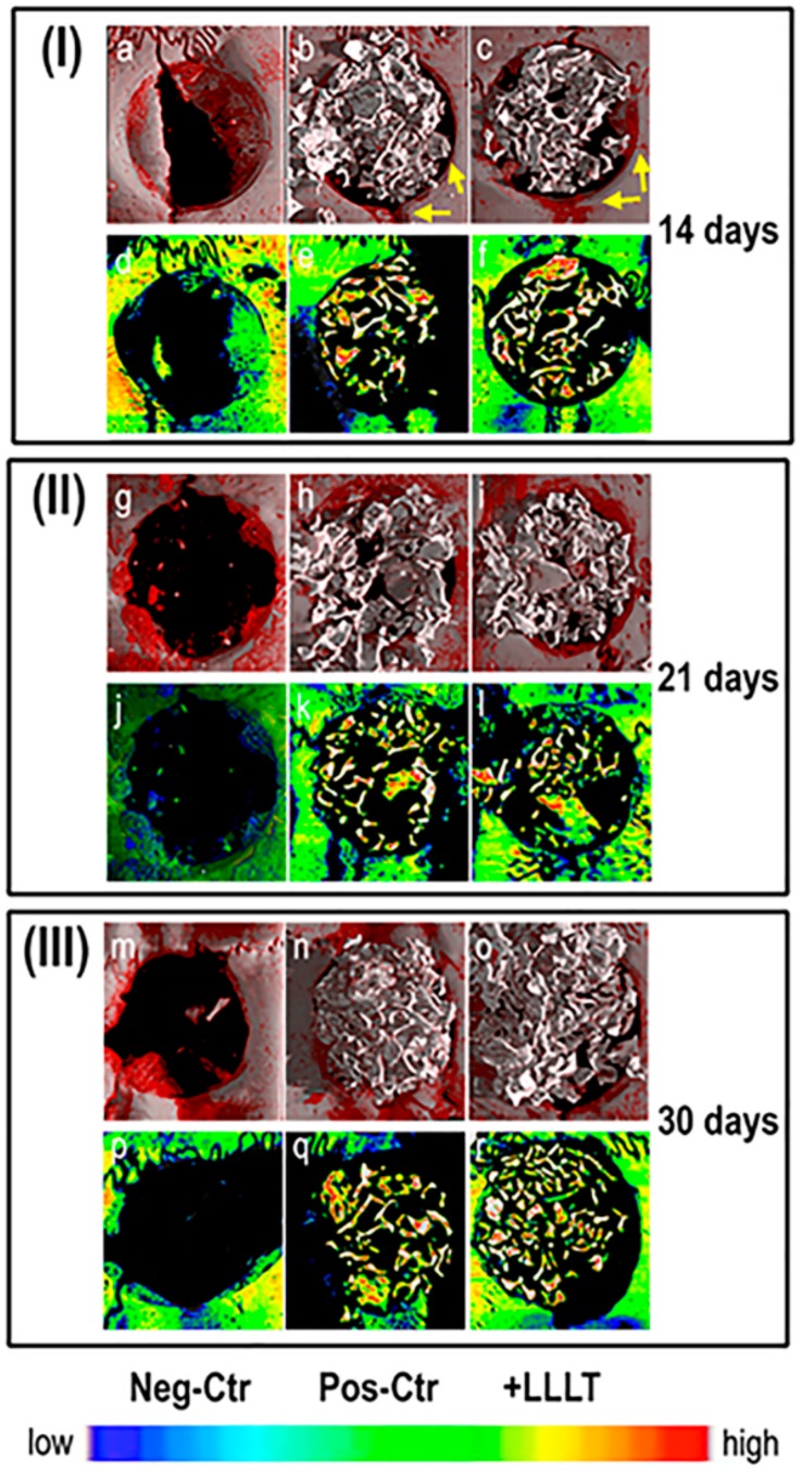
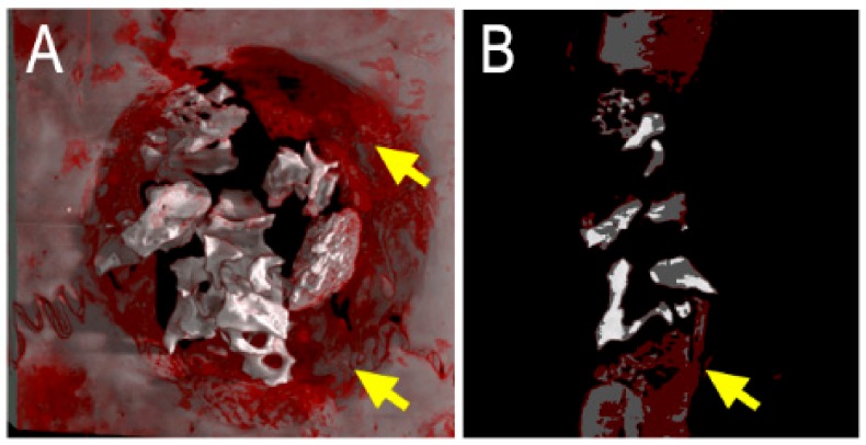
Background: Alveolar bone defects are usually the main concern when planning implant treatments for the appropriate oral rehabilitation of patients. To improve local conditions and achieve implant treatments, there are several methods used for increasing bone volume, among which one of the most successful, versatile, and effective is considered to be guided bone regeneration. The aim of this demonstrative study was to propose an innovative analysis protocol for the evaluation of the effect of photobiomodulation on the bone regeneration process, using rat calvarial defects of 5 mm in diameter, filled with xenograft, covered with collagen membrane, and then exposed to laser radiation.

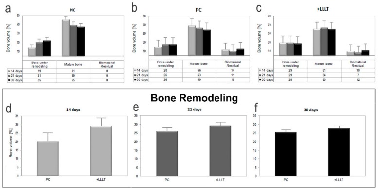
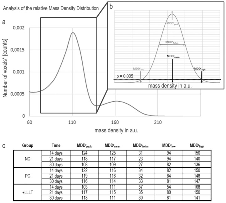
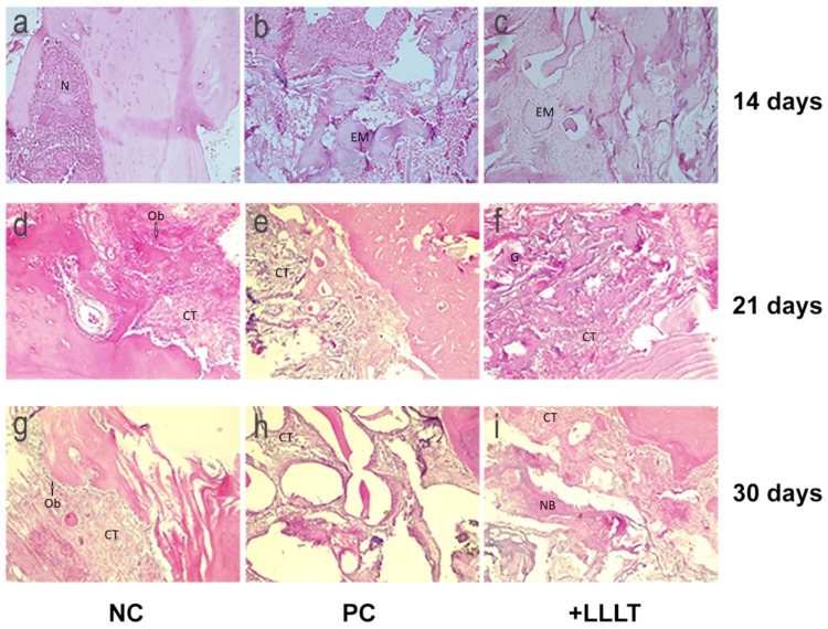
Methods: The animals were sacrificed at different points in time (i.e., after 14, 21, and 30 days). Samples of identical dimensions were harvested in order to compare the results obtained after different periods of healing. The analysis was performed by cross-linking the information obtained using histology and high-resolution synchrotron-based tomography on the same samples. A comparison was made with both the negative control (NC) group (with a bone defect which was left for spontaneous healing), and the positive control (PC) group (in which the bone defects were filled with xenografts and collagen membrane without receiving laser treatment).

Results: We demonstrated that using photobiomodulation provides a better healing effect than when receiving only the support of the biomaterial. This effect has been evident for short times treatments, i.e., during the first 14 days after surgery.

Conclusion: The proposed analysis protocol was effective in detecting the presence of higher quantities of bone volumes under remodeling after photobiomodulation with respect to the exclusive bone regeneration guided by the xenograft.

Keywords: Photobiomodulation; bone regeneration; collage membrane; histology; synchrotron radiation-based X-ray microtomography; xenograft.


Conflict of interest statement

The authors declare no conflict of interest.

Figures

Figure 1
Figure 1
Micro-CT images of repaired sites in representative retrieved samples. (af) Group I: samples harvested after 14 days: (ac) 3D reconstructions: (a) negative control (NC); (b) positive control (PC); (c) treated with low-level laser therapy (LLLT); (df) transversal sections of the defect: (d) NC; (e) PC; (f) treated with LLLT. (gl) Group II: samples harvested after 21 days: (gi) 3D reconstructions: (g) NC; (h) PC; (i) treated with LLLT; (jl) transversal sections of the defect: (j) NC; (k) PC; (l) treated with LLLT. (mr) Group III: samples harvested after 30 days: (mo) 3D reconstructions: (m) NC; (n) PC control; (o) treated with LLLT; (pr) transversal sections of the defect: (p) NC; (q) PC; (r) treated with LLLT. In 3D reconstructions grey tissue is mature bone; red tissue is bone under remodeling; white tissue is xenograft biomaterial; yellow arrows point to newly formed bone on defect border. In the transversal sections of the defect: white tissue is xenograft biomaterial; colors represent mineralization of the bone proportional to the color map in the bottom. Color map: blue stands for low mass density; red stands for high mass density.
Figure 2
Figure 2
Quantitative morphometric analysis. (ac) Mean volume percentages (vol.%) of the different mineralized phases (bone under remodeling, mature bone, and xenograft biomaterial) with respect to the overall mineralized volume: (a) NC group; (b) PC group; (c) +LLLT group. (df) Quantitative volumetric analysis of bone under remodeling portion, after (d) 14 days, (e) 21 days, and (f) 30 days from the surgery. Error bars are indicated.
Figure 3
Figure 3
Study of the relative mass density distribution (MDDr). (a) Portion of the histogram of a sampling biopsy: the peak on the left refers to the overall mineralized bone, the peak on the right refers to the biomaterial (xenograft) used to fill the defect; (b) study of the mineralized bone: the parameters investigated with the Roschger approach [38] are indicated. The threshold of p = 0.005 has been selected, as a good compromise to maintain a good sensitivity and minimize at the same time potential artifacts due to partial volume effects in the evaluation of MDDrlow; (c) parameters that derive from the profile fitting are indicated.
Figure 4
Figure 4
Histologic analysis. (a–c) Samples after 14 days of healing: (a) group NC, (b) group PC, and (c) group +LLLT. (df) Samples after 21 days of healing: (d) NC group, (e) PC group, and (f) +LLLT group. (gi) Samples after 30 days of healing: (g) NC group, (h) PC group, and (i) +LLLT group. N-necrosis; EM-eosinophilic material; CT-connective tissue; G-granulomas; Ob-osteoblasts. HE staining, original magnification: 10×.
Figure 5
Figure 5
Micro-CT images of repaired sites in representative retrieved sample, evaluated at 30 days postoperatively. The borders of the defect have not been covered with bovine bone graft, thus being directly exposed to laser radiation. A great amount of newly-formed bone can be observed at the periphery of the defect, as indicated with yellow arrows. (A): 3D reconstruction; (B): transversal section. Grey tissue is mature bone; red tissue is bone under remodeling; white tissue is xenograft biomaterial.

References

    1. Paknejad M., Rokn A., Rouzmeh N., Heidari M., Titidej A., Kharazifard M.J., Mehrfard A. Histologic evaluation of bone healing capacity following application of inorganic bovine bone and a new allograft material in rabbit calvaria. J. Dent. (Tehran) 2015;12:31–38. - PMC - PubMed
    1. Clokie C.M., Moghadam H., Jackson M.T., Sandor G.K. Closure of critical sized defects with allogenic and alloplastic bone substitutes. J. Craniofac. Surg. 2002;13:111–121. doi: 10.1097/00001665-200201000-00026. discussion 122–123. - DOI - PubMed
    1. Barboza E.P., Duarte M.E., Geolas L., Sorensen R.G., Riedel G.E., Wikesjo U.M. Ridge augmentation following implantation of recombinant human bone morphogenetic protein-2 in the dog. J. Periodontol. 2000;71:488–496. doi: 10.1902/jop.2000.71.3.488. - DOI - PubMed
    1. Wang W., Yeung K.W.K. Bone grafts and biomaterials substitutes for bone defect repair: A review. Bioact. Mater. 2017;2:224–247. doi: 10.1016/j.bioactmat.2017.05.007. - DOI - PMC - PubMed
    1. Yamada M., Egusa H. Current bone substitutes for implant dentistry. J. Prosthodont. Res. 2018;62:152–161. doi: 10.1016/j.jpor.2017.08.010. - DOI - PubMed