. 2020 May 11;21(5):1875-1885.
doi: 10.1021/acs.biomac.9b01745. Epub 2020 Feb 11.

Three-Dimensional Printed Cell Culture Model Based on Spherical Colloidal Lignin Particles and Cellulose Nanofibril-Alginate Hydrogel

Affiliations

Three-Dimensional Printed Cell Culture Model Based on Spherical Colloidal Lignin Particles and Cellulose Nanofibril-Alginate Hydrogel

Xue Zhang et al. Biomacromolecules. .

Abstract

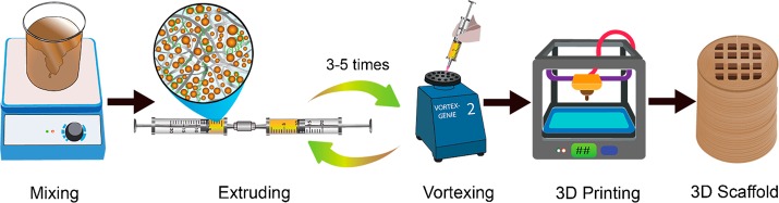
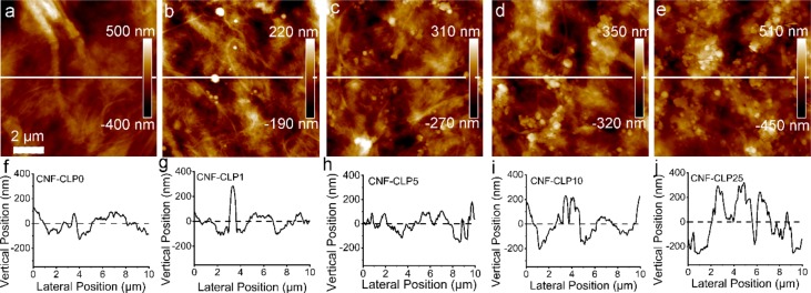
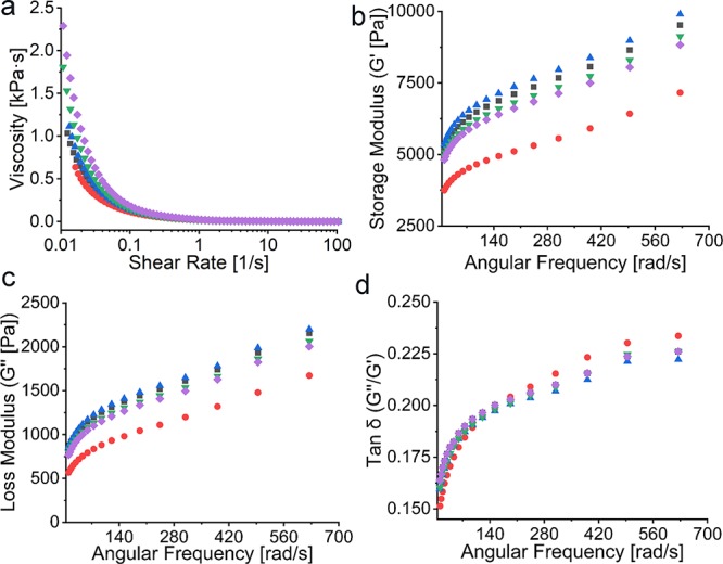
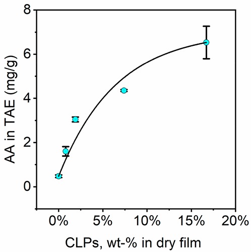
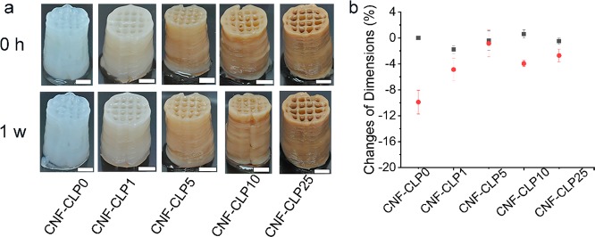
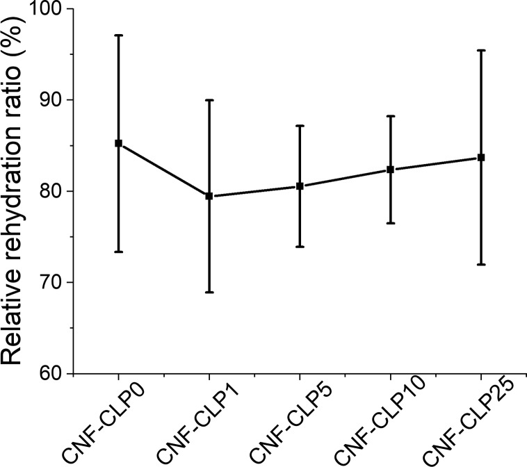
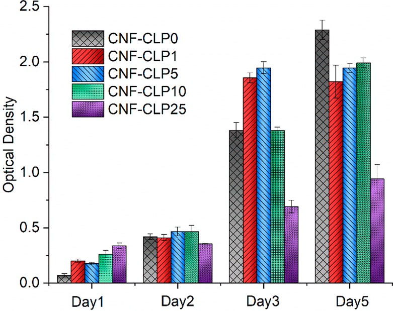
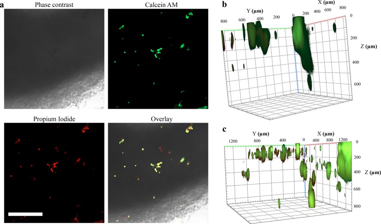
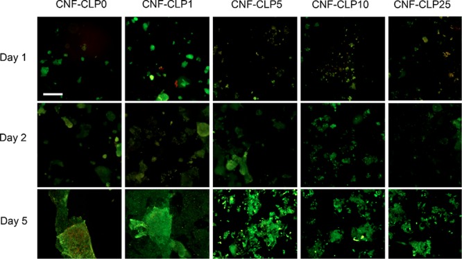
Three-dimensional (3D) printing has been an emerging technique to fabricate precise scaffolds for biomedical applications. Cellulose nanofibril (CNF) hydrogels have attracted considerable attention as a material for 3D printing because of their shear-thinning properties. Combining cellulose nanofibril hydrogels with alginate is an effective method to enable cross-linking of the printed scaffolds in the presence of Ca2+ ions. In this work, spherical colloidal lignin particles (CLPs, also known as spherical lignin nanoparticles) were used to prepare CNF-alginate-CLP nanocomposite scaffolds. High-resolution images obtained by atomic force microscopy (AFM) showed that CLPs were homogeneously mixed with the CNF hydrogel. CLPs brought antioxidant properties to the CNF-alginate-CLP scaffolds in a concentration-dependent manner and increased the viscosity of the hydrogels at a low shear rate, which correspondingly provide better shape fidelity and printing resolution to the scaffolds. Interestingly, the CLPs did not affect the viscosity at high shear rates, showing that the shear thinning behavior typical for CNF hydrogels was retained, enabling easy printing. The CNF-alginate-CLP scaffolds demonstrated shape stability after printing, cross-linking, and storage in Dulbecco's phosphate buffer solution (DPBS +) containing Ca2+ and Mg2+ ions, up to 7 days. The 3D-printed scaffolds showed relative rehydration ratio values above 80% after freeze-drying, demonstrating a high water-retaining capability. Cell viability tests using hepatocellular carcinoma cell line HepG2 showed no negative effect of CLPs on cell proliferation. Fluorescence microscopy indicated that HepG2 cells grew not only on the surfaces but also inside the porous scaffolds. Overall, our results demonstrate that nanocomposite CNF-alginate-CLP scaffolds have high potential in soft-tissue engineering and regenerative-medicine applications.


Conflict of interest statement

The authors declare no competing financial interest.

Figures

Scheme 1
Scheme 1. Schematic Illustration of Biomaterial Ink Preparation and 3D Printing
Note: not drawn to scale.
Figure 1
Figure 1
AFM height images of dry films of (a) CNF-CLP0, (b) CNF-CLP1, (c) CNF-CLP5, (d) CNF-CLP10, (e) CNF-CLP25, and the corresponding cross-section profiles (f–j). The cross-sectional profiles correspond to the white lines in (a–e) respectively. Scale bar, 2 μm.
Figure 2
Figure 2
Rheology data of CNF-CLP0 (■ black), CNFCLP1 (● red), CNF-CLP5 (▲ blue), CNF-CLP10 (▼ green), and CNF-CLP25 (◆ purple): (a) Dynamic viscosity curves of all the biomaterial ink formulations with shearing rate ranging from 0 to 100 1/s; (b) storage modulus G′ and (c) loss modulus G′′ of all the biomaterial inks formulations; (d) Tan δ (G′′/G′).
Figure 3
Figure 3
Antioxidant activity test of films produced from all CNF-alginate-CLP formulations with varying concentration of CLPs. Theoretical concentrations of CLPs in dry films were taken as X axis values.
Figure 4
Figure 4
(a) The printed scaffolds immediately after cross-linking (0 h) and after storage in DPBS+ (1 week). Scale bars are 0.5 cm. (b) The corresponding dimensional change ratio in height (■ black) and cross-section (● red) of printed scaffolds were measured immediately after cross-linking and after 1 week of storage in DPBS+. The dimension changes were calculated for two scaffolds, and the error bars represent the average of the absolute deviations relative to the mean value (N = 2). All the scaffolds were printed into a cylinder shape with a diameter of 1.5 cm and a height of 2 cm.
Figure 5
Figure 5
Relative rehydration ratios of freeze-dried scaffolds from different formulations, obtained through dividing the weight of rewetted scaffolds by the weight of the initial printed wet scaffolds. For each formulation, four scaffolds were measured, and the mean values were plotted. All the scaffolds were printed into a cylinder shape with a diameter of 8 mm and a height of 3 mm.
Figure 6
Figure 6
Biocompatibility test and HepG2 proliferation on scaffolds on day 1, day 2, day 3, and day 5.
Figure 7
Figure 7
HepG2 cells inside CNF-alginate-CLPs based printed scaffolds. (a) Microscopy images of CNF-CLP25 scaffolds after 1 day of incubation. Phase-contrast image, focused on the edge of the scaffold, displays the poor light permeability of the scaffolds. However, Calcein AM and PI staining enabled monitoring of cellular growth inside the scaffolds. Scale bar is 200 μm. (b) 3D reconstruction from Calcein AM and PI staining Z-stacks (5 μm spacing) of a CNF-CLP25 scaffold after 1 day of incubation, which displays the attachment of HepG2 cells mostly in a horizontal direction. (c) 3D reconstruction from Calcein AM and PI staining Z-stacks (5 μm spacing) of a CNF-CLP25 scaffold after 5 days of incubation, which displays the growth of HepG2 cells in a horizontal and vertical direction.
Figure 8
Figure 8
Fluorescence microscopy images of HepG2 cells, seeded on the formulated scaffolds, after 1 day, 2 days, and 5 days of incubation, with increasing relative concentrations of lignin to dry CNF (from 0 to 25%). Scale bar, 100 μm.

Similar articles

Cited by

References

    1. Chun H. J.; Park K.; Kim C.-H.; Khang G.. Novel Biomaterials for Regenerative Medicine; Springer, Singapore: Singapore, 2018.
    1. Tibbitt M. W.; Anseth K. S. Hydrogels as Extracellular Matrix Mimics for 3D Cell Culture. Biotechnol. Bioeng. 2009, 103, 655–663. 10.1002/bit.22361. - DOI - PMC - PubMed
    1. Gaspar V. M.; Lavrador P.; Borges J.; Oliveira M. B.; Mano J. F. Advanced Bottom-up Engineering of Living Architectures. Adv. Mater. 2019, 1903975.10.1002/adma.201903975. - DOI - PubMed
    1. Caliari S. R.; Burdick J. A. A Practical Guide to Hydrogels for Cell Culture. Nat. Methods 2016, 13 (5), 405–414. 10.1038/nmeth.3839. - DOI - PMC - PubMed
    1. De France K. J.; Badv M.; Dorogin J.; Siebers E.; Panchal V.; Babi M.; Moran-Mirabal J.; Lawlor M.; Cranston E. D.; Hoare T. Tissue Response and Biodistribution of Injectable Cellulose Nanocrystal Composite Hydrogels. ACS Biomater. Sci. Eng. 2019, 5 (5), 2235–2246. 10.1021/acsbiomaterials.9b00522. - DOI - PubMed

Publication types