. 2020 Mar 17;141(11):916-930.
doi: 10.1161/CIRCULATIONAHA.119.041882. Epub 2020 Jan 29.

Altering Sphingolipid Metabolism Attenuates Cell Death and Inflammatory Response After Myocardial Infarction

Affiliations

Altering Sphingolipid Metabolism Attenuates Cell Death and Inflammatory Response After Myocardial Infarction

Yoav Hadas et al. Circulation. .

Abstract

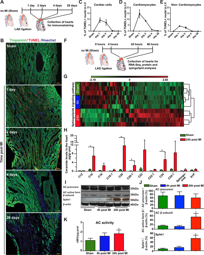
Background: Sphingolipids have recently emerged as a biomarker of recurrence and mortality after myocardial infarction (MI). The increased ceramide levels in mammalian heart tissues during acute MI, as demonstrated by several groups, is associated with higher cell death rates in the left ventricle and deteriorated cardiac function. Ceramidase, the only enzyme known to hydrolyze proapoptotic ceramide, generates sphingosine, which is then phosphorylated by sphingosine kinase to produce the prosurvival molecule sphingosine-1-phosphate. We hypothesized that Acid Ceramidase (AC) overexpression would counteract the negative effects of elevated ceramide and promote cell survival, thereby providing cardioprotection after MI.

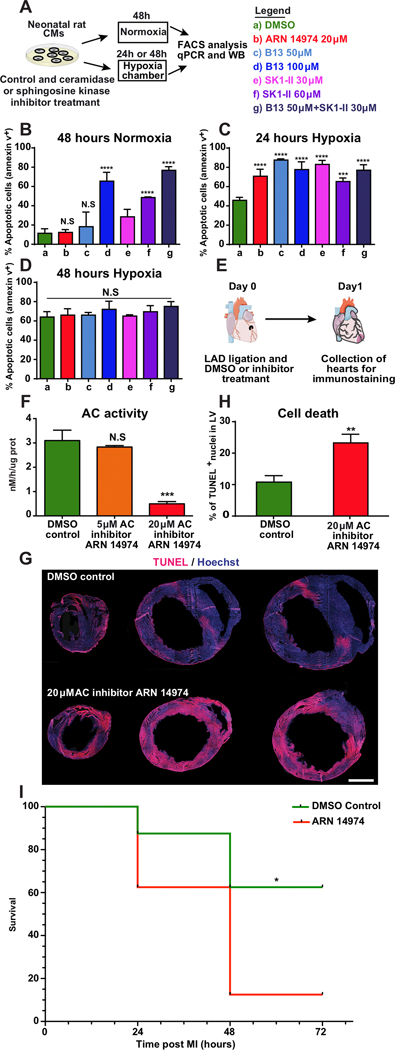
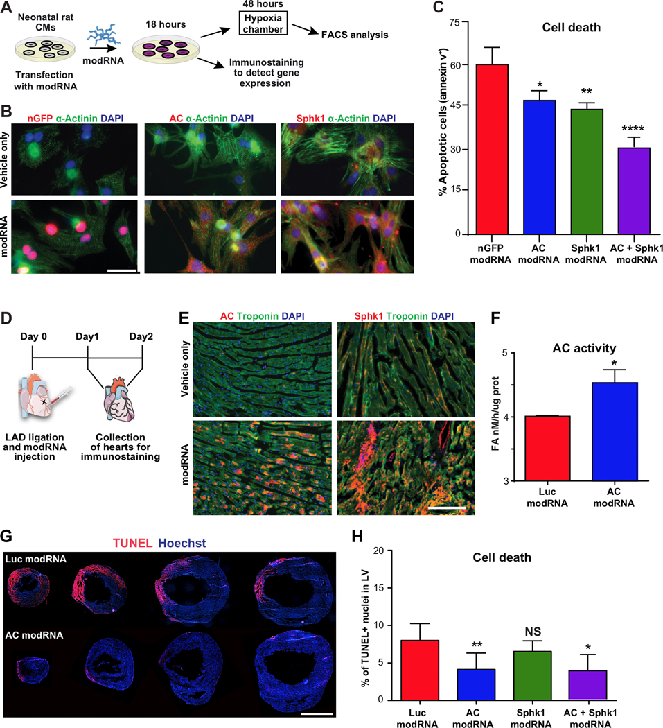
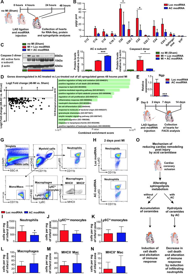
Methods: We performed transcriptomic, sphingolipid, and protein analyses to evaluate sphingolipid metabolism and signaling post-MI. We investigated the effect of altering ceramide metabolism through a loss (chemical inhibitors) or gain (modified mRNA [modRNA]) of AC function post hypoxia or MI.

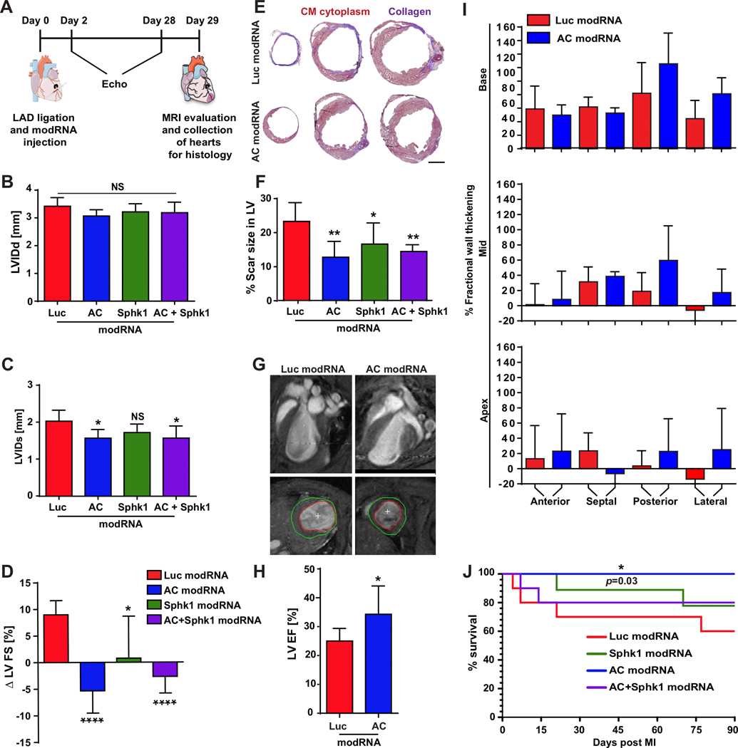
Results: We found that several genes involved in de novo ceramide synthesis were upregulated and that ceramide (C16, C20, C20:1, and C24) levels had significantly increased 24 hours after MI. AC inhibition after hypoxia or MI resulted in reduced AC activity and increased cell death. By contrast, enhancing AC activity via AC modRNA treatment increased cell survival after hypoxia or MI. AC modRNA-treated mice had significantly better heart function, longer survival, and smaller scar size than control mice 28 days post-MI. We attributed the improvement in heart function post-MI after AC modRNA delivery to decreased ceramide levels, lower cell death rates, and changes in the composition of the immune cell population in the left ventricle manifested by lowered abundance of proinflammatory detrimental neutrophils.

Conclusions: Our findings suggest that transiently altering sphingolipid metabolism through AC overexpression is sufficient and necessary to induce cardioprotection post-MI, thereby highlighting the therapeutic potential of AC modRNA in ischemic heart disease.

Keywords: acid ceramidase; cardioprotective agents; mRNA; myocardial infarction; sphingolipids.


Conflict of interest statement

CONFLICT OF INTEREST DISCLOSURES

E.E., L.Z, A.S.V. and Y.H. are Inventors on Provisional Patent Application (MODRNA ENCODING SPHINGOLIPID METABOLIZING PROTEINS TO PROMOTE CELL SURVIVAL) 3710/039P, Filed March 2018, which covers the results in this manuscript.

Figures

Figure 1.
Figure 1.. Characterizing cell death dynamics and sphingolipid metabolism in mouse hearts after MI.
A, Hearts were harvested from sham-operated mice or 1, 2, 4 and 28 days post MI. B, TUNEL assays were performed to assess DNA fragmentation in hearts harvested from either sham-operated mice or 1, 2, 4 or 28 days post MI. Troponin-I immunostaining was used to distinguish between cardiomyocytes and non-cardiomyocytes. Percentage of dead cells in left ventricle at day 1, 2, 4 and 28 post MI was quantified within all cardiac cells (DAPI+ cells) n=3 (C); CMs only (DAPI+, Troponin I+) n=3 (D) and non-CM cells only (DAPI+, Troponin I-) n=3 (E). F, For RNASeq, protein analysis and mass spectrometry, hearts were harvested from sham-operated mice or 4 or 24 hours post MI. RNA, proteins and lipids were extracted for sphingolipid determination. G, Hierarchical clustering dendrogram for the sphingolipid signaling pathway transcriptome in sham-operated hearts and hearts harvested 4 or 24 hours post MI, n=3, 3, 4, respectively. H, Sphingolipid levels measured in the LV of sham-operated mice or 24 hours post MI, n=3. I, Western blot of the AC precursor, AC active subunit β and Sphk1 in the LV of sham-operated hearts and hearts harvested 4 or 24 hours post MI. J, Quantified protein levels of AC precursor, AC β subunit and Sphk1, n=4. K, HPLC-MS/MS determination of AC activity in the LV of sham-operated hearts and hearts harvested 4 or 24 hours post MI, n=3. *, P<0.05, One-way ANOVA, Tukey’s Multiple Comparison Test for (J&K) and Holm-Sidak correction for multiple comparisons (H). Scale bar = 50μm. The results include two independent experiments for H-K.
Figure 2.
Figure 2.. Inhibiting sphingolipid metabolism increases cell death in cardiac cells in vitro and after MI.
A, Primary cardiomyocytes were isolated from the hearts of two- to three-day-old rats. Two days after isolation, the cells were treated with ceramidase inhibitors (pan-ceramidase inhibitor B13 and AC-specific ARN 14974) or sphingosine kinase inhibitor (SK1-II) and transferred either to normoxia (21% oxygen) for 48 hours or hypoxia (<2% oxygen) for 24 or 48 hours. At the end of the incubation period, the cells were stained with an apoptosis marker (Annexin 5) and a cell viability marker (DAPI) to assess the effects of inhibitors and oxygen levels on cell death. Cell death was quantified in cells treated with chemical inhibitors under normoxia for 48 hours, n=4 (B), or hypoxia for 24 hours, n=4 (C) and 48 hours, n=4 (D). E, AC inhibitor was injected intraperitoneally at the time of MI and 7 hours post MI. Hearts were harvested 24 hours post MI. Proteins were extracted for AC activity assays, or tissue was fixed and stained with TUNEL to assess cell death in the LV. F, HPLC-MS/MS determined AC activity in LV lysates 24 hours post MI and treatment with various AC inhibitor concentrations, n=3. G, Representative images of heart sections from mice treated with AC inhibitor or DMSO (control) obtained 24 hours post MI. H, Quantified cell death levels in the LV after treatment with AC inhibitor or DMSO control, 24 hours post MI, n=4. I, Short-term post MI survival curve for mice injected with AC inhibitor or DMSO control (n=8). ****, P<0.0001,***, P<0.001,**, P<0.01, *, P<0.05, NS, not significant. One-way ANOVA, Tukey’s Multiple Comparison Test (B-D&F), two-tailed Student’s t-test (H), Mantel-Cox log-rank test (I). Scale bar 1mm.
Figure 3.
Figure 3.. Acid ceramidase overexpression using modRNA decreases cell death in cardiac cells in vitro and after MI.
A, Primary cardiomyocytes were isolated from the hearts of two- to three-day-old rats. Two days after isolation, the cells were transfected with modRNAs for AC and SphK1. B, 18 hours post transfection in normoxia, CMs were fixed and immunostained to confirm modRNA translation. C, Cell death levels of CMs transfected with the nGFP modRNA (control), AC modRNA and SphK1 modRNA after 48 hours under hypoxia, n=4. D, modRNAs for Luc, AC and Sphk1 were injected into mouse hearts at the time of MI. To evaluate injected modRNA’s effects on the left ventricle, immunostaining (24 hours post MI), AC activity (24 hours post MI) and TUNEL assays were performed. E, Immunostaining for AC and SphK1 24 hours post modRNA injection. F, HPLC-MS/MS measurement of AC activity in the LV 24 hours post modRNA injection and MI, n=3 . G-H, Quantification and representative images of cell death levels in heart sections from mice treated with Luc modRNA (control), AC modRNA, Sphk1 modRNA or combination of AC + Sphk1 modRNAs 48 hours post injection, n=7. ****, P<0.0001, **, P<0.01, *, P<0.05, NS, not significant. One-way ANOVA, Tukey’s Multiple Comparison Test (C & H), Two-tailed Student’s t-test (F). Scale bars; 10μm (B), 50μm (E) and 1mm (G).
Figure 4.
Figure 4.. AC modRNA improves heart function and mouse survival post MI.
A, modRNAs for Luc, AC, Sphk1 or a combination of modRNAs for AC and Sphk1 were injected into the LV immediately after MI. Heart function was assessed by echocardiography and MRI. At day 29 post MI, hearts were collected for histological analysis. B-C, Echochardiography to measure left ventricular internal dimension end diastole (LVIDd) and left ventricular internal dimension end systole (LVIDs) 28 days post MI, n=6. D, Echocardiography to determine changes in left ventricle fractional shortening (FS) between day 2 and day 28 post MI, n=6 . E, Representative images of Masson’s trichrome staining and F, percentage of left ventricle area occupied by scar tissue 28 days post MI and modRNA injection, n=6. G-H, Representative images of MRI scans and quantification of left ventricle ejection fraction (LVEF) 29 days post MI and injection with AC or Luc modRNA. I, MRI-based measurement of fractional wall thickening percentage 29 days post MI and injection with AC or Luc modRNA, n=6. J, Long-term survival of mice after MI and transfection with different modRNAs, n=10. ****, P<0.0001, **, P<0.01, *, P<0.05, NS, not significant. One-way ANOVA, Bonferroni post-hoc tests (B, C, D, F) and two-tailed Student’s t-tests (H). The results include two independent experiments. Scale bar = 1mm.
Figure 5.
Figure 5.. AC modRNA reduces ceramide levels and alters the immune cell composition in the left ventricle post MI.
A, modRNAs for Luc or AC were injected into mouse hearts immediately after MI. Lipids and proteins were extracted from the hearts 24 hours post MI to analyze sphingolipids by mass spectrometry and proteins by western blot. For RNA sequencing, hearts were harvested 4, 24 or 48 hours post MI. B, Sphingolipid levels after Luc or AC modRNA treatment, assessed 24 hours post MI, n=3. C, Western blot and quantification of AC α subunits and caspase 3 dimers in sham-operated hearts and in hearts treated with Luc or AC modRNA, 24 hours post MI, n=3. D, Scatterplot and GO enrichment analysis of all genes downregulated in AC-treated vs. Luc-treated out of all upregulated genes 2 days post MI, n=4. E, Relative Ngp gene expression levels 2 days post MI in the LV of mice treated with Luc or AC, quantified using qPCR, n=4. F, To analyze modRNA treatment’s effect on immune cell content after MI, hearts were collected at 2, 7 and 14 days post MI and Luc or AC modRNA injection and analyzed using flow cytometry. G, Gating strategy for immune cell populations in the infarct zone of modRNA-treated hearts. Neutrophils are defined as CD45+ CD11b+ Ly6G+, macrophages as CD45+ CD11b+ Lin- F4/80+ Ly6Clow/int and monocytes as CD45+ CD11b+ Lin- F4/80- Ly6Clow / Ly6Chi. H, Representative plots of neutrophil populations in Luc modRNA or AC modRNA-treated hearts 2 days post MI. I-N, Flow cytometric quantification of immune cell subsets in the infarct zone of Luc modRNA- and AC modRNA-treated hearts 2 days post MI included following populations: neutrophils, n=16 (I); Ly6Clow monocytes, n=16 (J); Ly6Chi monocytes, n=16 (K); total macrophages, n=16 (L), MHCII-, n=16 (M) and MHCII+, n=16 (N) macrophage subsets, n=16. O, Summary of proposed AC modRNA action mechanism during and after acute MI. ***, P<0.001, *, P<0.05, n=3 (B, C), n=4 (E), n=16–18 (I-N). Holm-Sidak correction for multiple comparisons (B), one-way ANOVA, Tukey’s Multiple Comparison Test (C) and two-tailed Student’s t-tests (E&I-N). Scale bar 20μm. The results include two and three (FACS) independent experiments.

References

    1. Spencer FA, Meyer TE, Gore JM and Goldberg RJ. Heterogeneity in the management and outcomes of patients with acute myocardial infarction complicated by heart failure: the National Registry of Myocardial Infarction. Circulation. 2002;105:2605–2610. - PubMed
    1. Hadas Y, Katz MG, Bridges CR and Zangi L. Modified mRNA as a therapeutic tool to induce cardiac regeneration in ischemic heart disease. Wiley Interdiscip Rev Syst Biol Med. 2017;9: wsbm.1367. - PMC - PubMed
    1. Lin Z and Pu WT. Strategies for cardiac regeneration and repair. Sci Transl Med. 2014;6:239rv1. - PMC - PubMed
    1. Meeusen JW, Donato LJ and Jaffe AS. Lipid Biomarkers for Risk Assessment in Acute Coronary Syndromes. Curr Cardiol Rep. 2017;19:48. - PubMed
    1. Yu J, Pan W, Shi R, Yang T, Li Y, Yu G, Bai Y, Schuchman EH, He X and Zhang G. Ceramide is upregulated and associated with mortality in patients with chronic heart failure. Can J Cardiol. 2015;31:357–363. - PubMed

Publication types

MeSH terms