. 2020 Jan 28;11(1):38.
doi: 10.1186/s13287-020-1562-9.

Exosomes from bone marrow mesenchymal stem cells enhance fracture healing through the promotion of osteogenesis and angiogenesis in a rat model of nonunion

Affiliations

Exosomes from bone marrow mesenchymal stem cells enhance fracture healing through the promotion of osteogenesis and angiogenesis in a rat model of nonunion

Lu Zhang et al. Stem Cell Res Ther. .

Abstract

Background: As important players in cell-to-cell communication, exosomes (exo) are believed to play a similar role in promoting fracture healing. This study investigated whether exosomes derived from bone marrow mesenchymal stem cells (BMMSC-Exos) could improve fracture healing of nonunion.

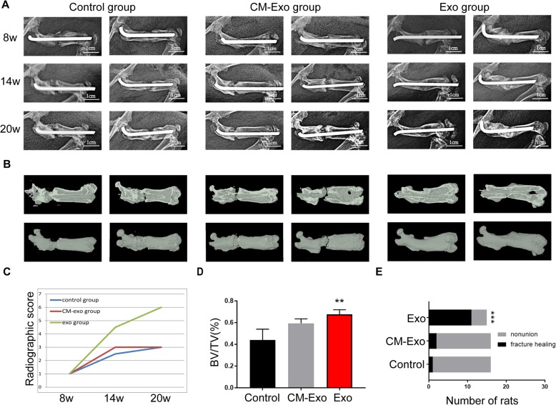
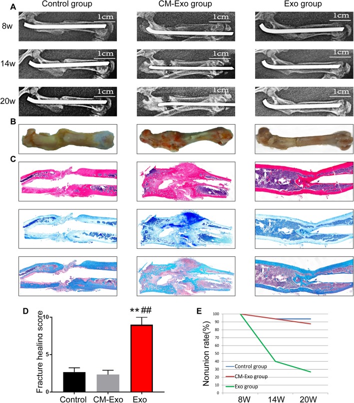
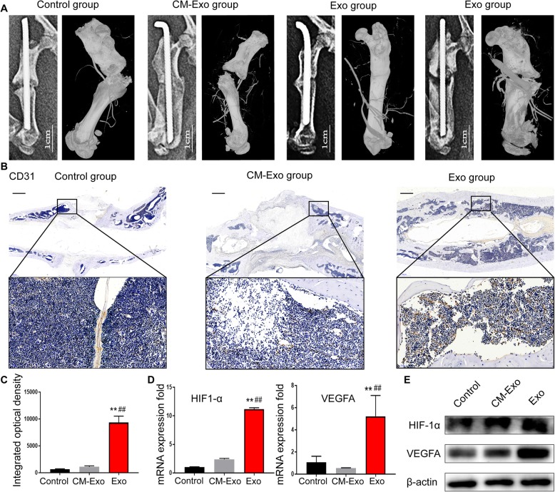
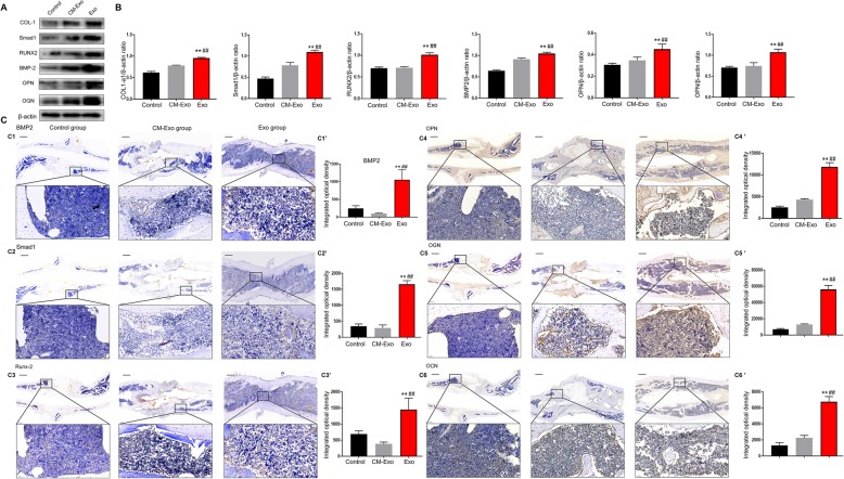
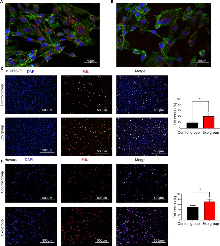
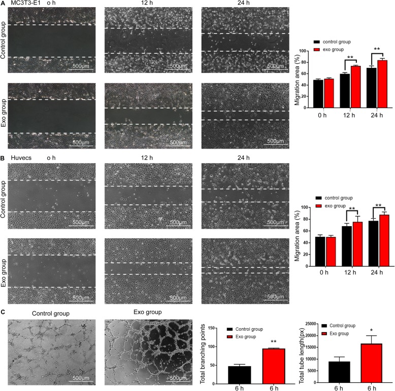
Methods: BMMSC-Exos were isolated and transplanted into the fracture site in a rat model of femoral nonunion (Exo group) every week. Moreover, equal volumes of phosphate-buffered saline (PBS) and exosome-depleted conditioned medium (CM-Exo) were injected into the femoral fracture sites of the rats in the control and CM-Exo groups. Bone healing processes were recorded and evaluated by radiographic methods on weeks 8, 14 and 20 after surgery. Osteogenesis and angiogenesis at the fracture sites were evaluated by radiographic and histological methods on postoperative week 20. The expression levels of osteogenesis- or angiogenesis-related genes were evaluated in vitro by western blotting and immunohistochemistry. The ability to internalize exosomes was assessed using the PKH26 assay. Altered proliferation and migration of human umbilical vein endothelial cells (HUVECs) and mouse embryo osteoblast precursor cells (MC3TE-E1s) treated with BMMSC-Exos were determined by utilizing EdU incorporation, immunofluorescence staining, and scratch wound assay. The angiogenesis ability of HUVECs was evaluated through tube formation assays. Finally, to explore the effect of exosomes in osteogenesis via the BMP-2/Smad1/RUNX2 signalling pathway, the BMP-2 inhibitors noggin and LDN193189 were utilized, and their subsequent effects were observed.

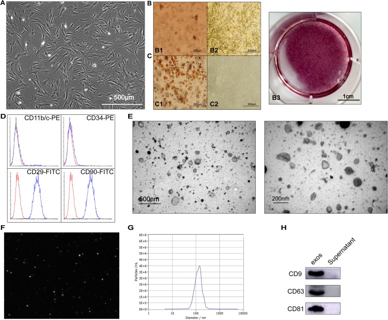
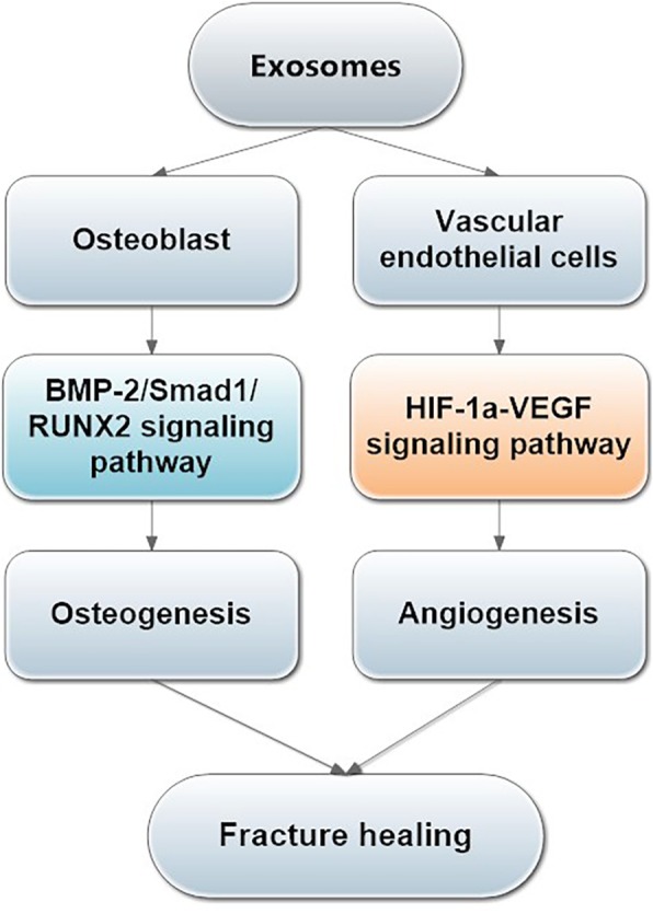
Results: BMMSC-Exos were observed to be spherical with a diameter of approximately 122 nm. CD9, CD63 and CD81 were expressed. Transplantation of BMMSC-Exos obviously enhanced osteogenesis, angiogenesis and bone healing processes in a rat model of femoral nonunion. BMMSC-Exos were taken up by HUVECs and MC3T3-E1 in vitro, and their proliferation and migration were also improved. Finally, experiments with BMP2 inhibitors confirmed that the BMP-2/Smad1/RUNX2 signalling pathway played an important role in the pro-osteogenesis induced by BMMSC-Exos and enhanced fracture healing of nonunion.

Conclusions: Our findings suggest that transplantation of BMMSC-Exos exerts a critical effect on the treatment of nonunion by promoting osteogenesis and angiogenesis. This promoting effect might be ascribed to the activation of the BMP-2/Smad1/RUNX2 and the HIF-1α/VEGF signalling pathways.

Keywords: Angiogenesis; Exosomes; Nonunion; Osteogenesis.


Conflict of interest statement

The authors declare that they have no competing interests.

Figures

Fig. 1
Fig. 1
Characterization of BMMSCs and BMMSC-Exos. a Fusiform morphology of BMMSCs shown in light microscopy images. b Alizarin red staining was performed to detect the osteogenic differentiation ability of BMMSCs: B1, staining of experimental group; B2, staining of control group; B3, gross scanning images of ARS staining of experimental group. c Oil red staining was performed to detect the lipid differentiation ability of BMMSCs: C1, staining of the experimental group; C2, staining of the control group. d Surface markers of BMMSCs analysed by flow cytometry. The cells were negative for CD34 and CD11b/C and positive for CD90 and CD29. e The morphology of BMMSC-Exos shown by TEM. f Image of the purified exosomes. g The particle size distribution in purified BMMSC-Exos determined by NTA. h The surface markers (CD9, CD63 and CD81) of exosomes were detected by western blotting
Fig. 2
Fig. 2
Radiographic analysis of fracture healing. a Representative radiological images of the control, CM-Exo and Exo groups at 8, 14 and 20 weeks after surgery. (Radiological examination 8 weeks after the surgery was performed to confirm successful nonunion modelling). b Representative micro-CT images of a nonunion femur at 20 weeks after surgery. c Radiographic score for X-ray images on postoperative weeks 8, 14 and 20, n = 5. d BV/TV on postoperative week 20 was quantified, n = 3. e Bone union rate was calculated by chi-square test in the control, CM-Exo and Exo groups. Abbreviations: 8w, 8 weeks; CM, conditioned medium; CM-Exo, exosome-depleted conditioned medium; Exo, exosomes; BV, bone volume; TV, total volume. **P < 0.01; ***P < 0.001
Fig. 3
Fig. 3
Radiographic and histological analysis of femoral specimens. a, b Radiographic imaging by X-ray was performed at 8, 14 and 20 weeks after the surgery, and rat femurs were harvested at 20 weeks after the surgery. c H&E, toluidine blue and safranin O-fast green staining of the femurs. d Quantification of fracture healing in calluses. e Bone nonunion rate of the control, CM-Exo and Exo groups. Abbreviations: 8w, 8 weeks; H&E, haematoxylin-eosin. **Statistically significant difference compared with the control group (P < 0.01). ##Statistically significant difference compared with the CM-Exo group (P < 0.01)
Fig. 4
Fig. 4
Assessment of angiogenesis at the fracture site. a Representative micro-CT images of the vasculature in the fracture site on postoperative week 20. b Anti-CD31 staining of the femur slices on postoperative week 20. Representative areas are shown, and boxed areas are enlarged on the bottom. Scale bar = 1 mm. c The integrated optical density of CD31-positive vessels was measured. d Total RNA was extracted, and the expression levels of osteogenesis-related genes (HIF1-α and VEGF) in the bone tissue of the fracture sites were analysed by qRT-PCR, n = 3. e Western blot analysis showed that HIF-1α and VEGF protein levels increased in the bone tissue of the fracture sites stimulated with BMMSC-Exos. Abbreviations: CM, conditioned medium; CM-Exo, exosome-depleted conditioned medium; Exo, exosomes. **P < 0.01 vs the control group; ##P < 0.01 vs the CM-Exo group
Fig. 5
Fig. 5
Assessment of osteogenesis in the bone tissue. a Western blot analysis showed that the Smad1, RUNX2, BMP-2, OPN and OGN protein levels increased in the bone tissue of the fracture sites stimulated with BMMSC-Exos. b The grey value ratios were quantified using ImageJ software (NIH, USA). The experiments were performed on three separate occasions. c The immunohistochemical analysis of BMMSC-Exos promoted osteogenesis in vivo. Immunohistochemical analysis of BMP-2 (C1), Smad1 (C2), RUNX2 (C3), OPN (C4), OGN (C5) and OCN (C6) was used to detect osteogenesis in the femur specimens. The brown colour represents positive staining of BMP-2, Smad1, RUNX2, OPN, OGN and OCN. The integrated optical density of positive staining was measured (C1′–C6′). Abbreviations: CM, conditioned medium; CM-Exo, exosome-depleted conditioned medium; Exo, exosomes. **P < 0.01 vs the control group; ##P < 0.01 vs the CM-Exo group
Fig. 6
Fig. 6
BMMSC-Exos entered MC3T3-E1Cs and HUVECs and promoted the proliferation of the cells. a, b Laser scanning confocal microscopy analysis of the internalization of PKH26-labelled BMMSC-Exos by MC3T3-E1Cs (a) and HUVECs (b). The red-labelled exosomes were visible in the perinuclear region of recipient cells. c, d EdU incorporation by the control and Exo groups of MC3T3-E1Cs (c) and HUVCs (d) was visualized using a fluorescence microscope. The percentage of EdU-positive cells for each group was quantitated using ImageJ software (right graph), n = 3. Abbreviation: Exo, exosomes. *P < 0.05 vs the control group
Fig. 7
Fig. 7
BMMSC-Exos enhanced the migration ability and angiogenesis of MC3T3-E1Cs and HUVECs. a, b Scratched wound assay and quantitative analysis of MC3T3-E1Cs and HUVECs. The migration area was significantly greater in the Exo group than in the control group at 12 h and 24 h. c BMMSC-Exos stimulated the tube formation ability of HUVECs, and quantitative analysis is shown in the graphs on the right. Abbreviation: Exo, exosomes. *P < 0.05 vs the control group; **P < 0.01 vs the Control group
Fig. 8
Fig. 8
The inhibition of BMP-2 decreased the Smad1 and RUNX2 protein levels in MC3T3-E1Cs stimulated with BMMSC-Exos. a Western blot analysis showed that even if BMMSC-Exos were applied, the expression levels of BMP-2, Smad1 and RUNX2 proteins were markedly increased after BMP-2 was inhibited. b The grey value ratios were quantified. c The immunocytochemistry analysis showed that applying BMMSC-Exos+BMP-2 inhibitor could not promote osteogenesis. d Gross scanning images of ALP and ARS staining of MC3T3-E1Cs (control group and BMMSC-Exos group), ALP staining at 14 days and ARS staining at 21 days. e Quantitative analysis of ALP and ARS activity was used to evaluate the effect of BMMSC-Exos on the osteogenic ability of MC3T3-E1Cs
Fig. 9
Fig. 9
Research schematic. BMMSC-Exos could accelerate the proliferation and migration of osteoblast cells and endothelial cells, further promoting angiogenesis and osteogenesis by activating the HIF-1α/VEGF and the BMP-2/Smad1/RUNX2 signalling pathways to enhance fracture healing

References

    1. Einhorn TA, Gerstenfeld LC. Fracture healing: mechanisms and interventions. Nat Rev Rheumatol. 2015;11(1):45. doi: 10.1038/nrrheum.2014.164. - DOI - PMC - PubMed
    1. Murata K, Ito H, Yoshitomi H, et al. Inhibition of miR-92a enhances fracture healing via promoting angiogenesis in a model of stabilized fracture in young mice. J Bone Miner Res. 2014;29(2):316–326. doi: 10.1002/jbmr.2040. - DOI - PubMed
    1. Ho-Shui-Ling A, Bolander J, Rustom LE, Johnson AW, Luyten FP, Picart C. Bone regeneration strategies: engineered scaffolds, bioactive molecules and stem cells current stage and future perspectives. Biomaterials. 2018;180:143. doi: 10.1016/j.biomaterials.2018.07.017. - DOI - PMC - PubMed
    1. Granero-Moltó F, Myers TJ, Weis JA, et al. Mesenchymal stem cells expressing insulin-like growth factor-I (MSCIGF) promote fracture healing and restore new bone formation in Irs1 knockout mice: analyses of MSCIGF autocrine and paracrine regenerative effects. Stem Cells. 2011;29(10):1537. doi: 10.1002/stem.697. - DOI - PMC - PubMed
    1. Wei CC, Lin AB, Hung SC. Mesenchymal stem cells in regenerative medicine for musculoskeletal diseases: bench, bedside, and industry. Cell Transplant. 2014;23(4–5):505. doi: 10.3727/096368914X678328. - DOI - PubMed