. 2020 Mar 13;295(11):3652-3663.
doi: 10.1074/jbc.RA119.011646. Epub 2020 Jan 28.

Rab35-GEFs, DENND1A and folliculin differentially regulate podocalyxin trafficking in two- and three-dimensional epithelial cell cultures

Affiliations

Rab35-GEFs, DENND1A and folliculin differentially regulate podocalyxin trafficking in two- and three-dimensional epithelial cell cultures

Riko Kinoshita et al. J Biol Chem. .

Abstract

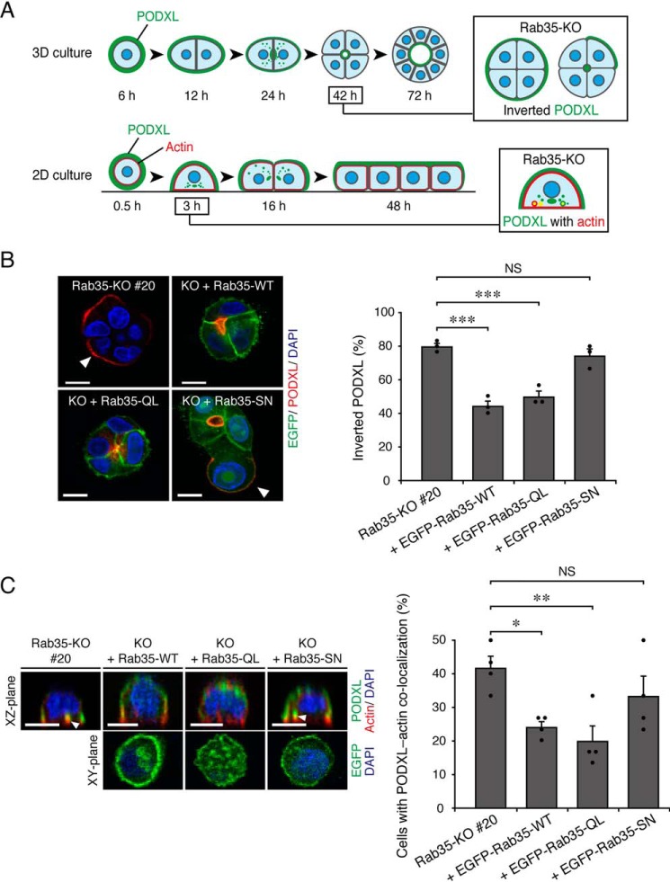
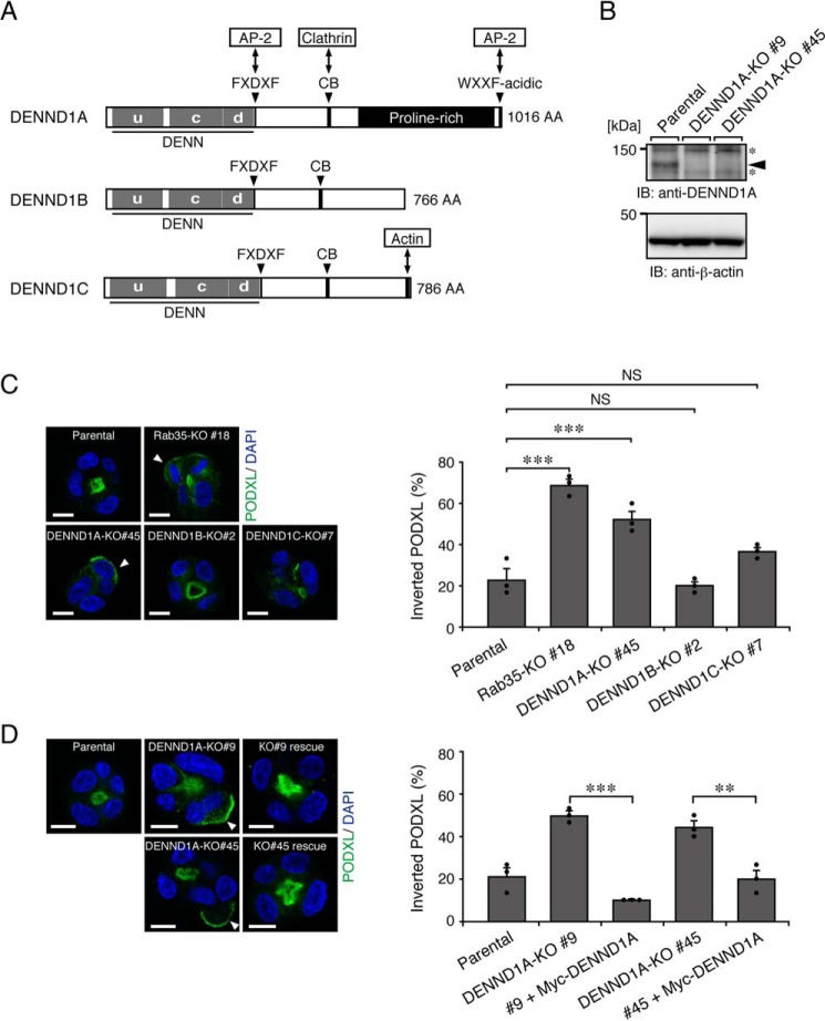
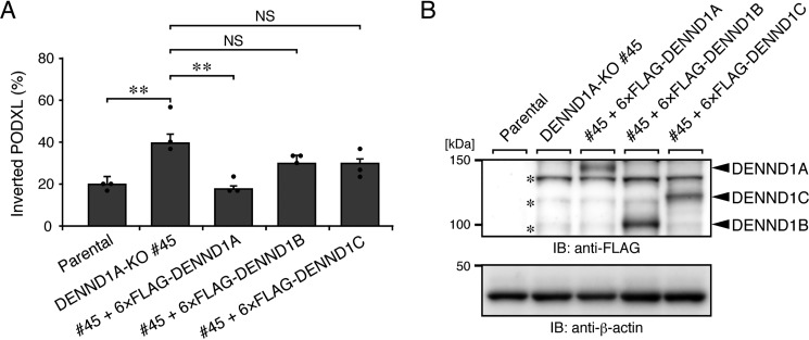
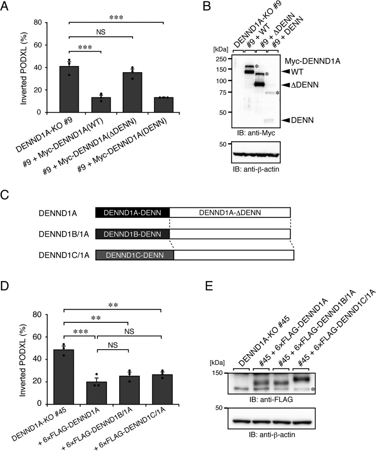
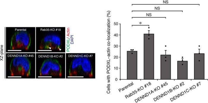
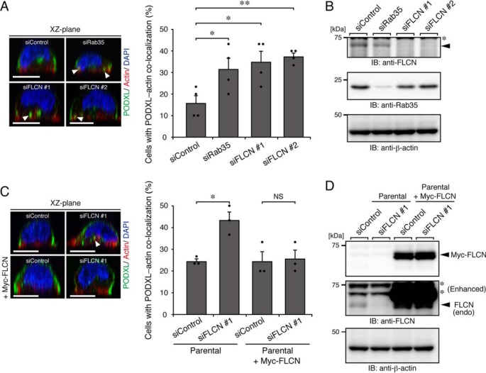
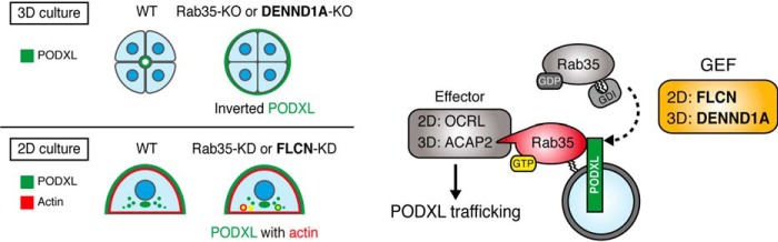
Polarized epithelial cells have functionally distinct apical and basolateral membranes through which they communicate with external and internal bodily environments, respectively. The establishment and maintenance of this asymmetric structure depend on polarized trafficking of specific cargos, but the precise molecular mechanism is incompletely understood. We previously showed that Rab35, a member of the Rab family small GTPases, differentially regulates the trafficking of an apical cargo, podocalyxin (PODXL), in two-dimensional (2D) and three-dimensional (3D) Madin-Darby canine kidney (MDCK) II cell cultures through specific interactions with two distinct effectors, OCRL inositol polyphosphate-5-phosphatase (OCRL) and ArfGAP with coiled-coil, ankyrin repeat and pleckstrin homology domains 2 (ACAP2), respectively. However, whether the upstream regulators of Rab35 also differ depending on the culture conditions remains completely unknown. Here, we investigated four known guanine nucleotide exchange factors (GEFs) of Rab35, namely DENN domain-containing 1A (DENND1A), DENND1B, DENND1C, and folliculin (FLCN), and demonstrate that DENND1A and FLCN exhibit distinct requirements for Rab35-dependent PODXL trafficking under the two culture conditions. In 3D cell cultures, only DENDN1A-knockout cysts exhibited the inverted localization of PODXL similar to that of Rab35-knockout cysts. Moreover, the DENN domain, harboring GEF activity toward Rab35, was required for proper PODXL trafficking to the apical membrane. By contrast, FLCN-knockdown cells specifically accumulated PODXL in actin-rich structures similar to the Rab35-knockdown cells in 2D cell cultures. Our findings indicate that two distinct functional cascades of Rab35, the FLCN-Rab35-OCRL and the DENND1A-Rab35-ACAP2 axes, regulate PODXL trafficking in 2D and 3D MDCK II cell cultures, respectively.

Keywords: 3D cell culture; Rab; cell polarity; epithelial cell; guanine nucleotide exchange factor (GEF); membrane trafficking; podocalyxin; small GTPase; tissue development.


Conflict of interest statement

The authors declare that they have no conflicts of interest with the contents of this article

Figures

Figure 1.
Figure 1.
Active Rab35 is required for PODXL trafficking under 2D and 3D culture conditions. A, schematic representation of PODXL localization during 3D cyst and 2D monolayer formation of MDCK II cells (modified from Ref. 6). In a 3D Rab35-KO cell culture, PODXL (green) remained on the outer membrane at 42 h after plating (Inverted PODXL) (top row, 3D culture). In a 2D Rab35-KO cell culture, endocytosed PODXL from the membrane attaching to the bottom plane of the dish localized and accumulated on actin-rich structures (red) at 3 h after plating (PODXL with actin) (bottom row, 2D culture). B, Rab35-KO (#20) and its rescued cells (+EGFP-Rab35 (WT, QL, or SN; shown in green)) were plated on Matrigel and fixed at 42 h after plating. The cells were stained for PODXL (red) and DAPI (blue), followed by counting of the inverted PODXL (30 cysts/condition). The arrowheads show PODXL localized on the outer membrane. Scale bars, 10 μm. The graph shows the means and S.E. (error bars) of three independent experiments. ***, p < 0.001; NS, not significant (Dunnett's test). C, Rab35-KO (#20) and its rescued cells (+EGFP-Rab35 (WT, QL, or SN)) were plated on glass-bottom dishes and fixed at 3 h after plating. The cells were stained for PODXL (green), actin (red), and DAPI (blue), followed by counting of cells with co-localized PODXL and actin (30 cells/condition). The confocal xz-plane (top) and the xy-plane (bottom) are shown. The arrowheads show PODXL co-localizing with actin. Scale bars, 10 μm. The graph shows the means and S.E. (error bars) of four independent experiments. *, p < 0.05; **, p < 0.01; NS, not significant (Dunnett's test).
Figure 2.
Figure 2.
DENND1A-KO, but not DENND1B-KO or DENND1C-KO, induces the inverted localization of PODXL in 3D cysts. A, schematic representation of mouse DENND1 family proteins. The domains and motifs are depicted according to previous reports (15, 17) and the UniProtKB (DENND1A, Q8K382; DENND1B, Q3U1T9; DENND1C, Q8CFK6). uDENN, upstream DENN; cDENN, central/core DENN; dDENN, downstream DENN; FXDXF, AP-2 α-ear platform subdomain–binding motif; CB, clathrin heavy chain–binding motif; WXXF-acidic, AP-2 α-ear sandwich subdomain–binding motif; AA, amino acids. B, lysates of parental cells and two DENND1A-KO cells (#9 and #45) were analyzed by immunoblotting (IB) with anti-DENND1A and anti-β-actin antibodies. The arrowhead indicates the position of endogenous DENND1A. The asterisks indicate nonspecific bands of the primary antibody. C, parental, Rab35-KO, DENND1A-KO, DENND1B-KO, and DENND1C-KO cells were plated on Matrigel and fixed at 42 h after plating. The cells were stained for PODXL (green) and DAPI (blue), followed by counting of the inverted PODXL (30 cysts/condition). The arrowheads show PODXL localizing on the outer membrane. Scale bars, 10 μm. The graph shows the means and S.E. (error bars) of three independent experiments. ***, p < 0.001; NS, not significant (Dunnett's test). D, parental cells, DENND1A-KO (#9 and #45) cells, and their rescued cells (+Myc-DENND1A) were plated on Matrigel and fixed at 42 h after plating, followed by counting of the inverted PODXL (30 cysts/condition). The arrowheads show PODXL localizing on the outer membrane. Scale bars, 10 μm. The graph shows the means and S.E.(error bars) of three independent experiments. ***, p < 0.001; **, p < 0.01 (Tukey's test).
Figure 3.
Figure 3.
DENND1A is specifically required for PODXL trafficking in 3D cysts. A, parental cells, DENND1A-KO (#45) cells, and its rescued cells (+6×FLAG-tagged DENND1A, DENND1B, or DENND1C) were plated on Matrigel and fixed at 42 h after plating, followed by counting of the inverted PODXL (30 cysts/condition). The graph shows the means and S.E. (error bars) of three independent experiments. **, p < 0.01; NS, not significant (Tukey's test). Representative images are shown in Fig. S3A. B, lysates of the cells used in A were analyzed by immunoblotting (IB) with anti-FLAG and anti-β-actin antibodies. The asterisks show the predicted 3×FLAG-tagged SpCas9 degradation products, because an ∼160-kDa protein was detected by immunoblotting with anti-FLAG antibody in DENND1A-KO #45 cells, but not in parental cells (data not shown).
Figure 4.
Figure 4.
The N-terminal DENN domain of DENND1A is required for PODXL trafficking in 3D cysts. A, DENND1A-KO (#9) and its rescued cells (+Myc-DENND1A (WT, ΔDENN, or DENN)) were plated on Matrigel and fixed at 42 h after plating, followed by counting of the inverted cysts (30 cysts/condition). The graph shows the means and S.E. (error bars) of three independent experiments. ***, p < 0.001; NS, not significant (Dunnett's test). Representative images are shown in Fig. S3B. B, lysates of the cells used in A were analyzed by immunoblotting (IB) with anti-Myc and anti-β-actin antibodies. The additional higher bands (asterisks) in the blots presumably result from post-translational modifications, such as phosphorylation (30). Alternatively, the DENN domain of DENND1A could form an SDS-insensitive dimer. C, schematic representation of DENND1B/1A and DENND1C/1A chimera proteins. The DENND1B/1A (or DENND1C/1A) chimera protein consists of the DENN domain of human DENND1B (DENND1B-DENN) (or DENND1C (DENND1C-DENN)) fused with the C-terminal part of mouse DENND1A (DENND1A-ΔDENN). D, DENND1A-KO (#45) and its rescued cells (+6×FLAG-tagged DENND1A, DENND1B/1A, or DENND1C/1A) were plated on Matrigel and fixed at 42 h after plating, followed by counting of the inverted PODXL (30 cysts/condition). The graph shows the means and S.E. (error bars) of three independent experiments. ***, p < 0.001; **, p < 0.01; NS, not significant (Tukey's test). Representative images are shown in Fig. S3C. E, lysates of the cells used in D were analyzed by immunoblotting with anti-FLAG and anti-β-actin antibodies. The asterisk shows the predicted 3×FLAG-tagged SpCas9 degradation products.
Figure 5.
Figure 5.
DENND1 family proteins are not required for PODXL trafficking under 2D culture conditions. Parental, Rab35-KO, DENND1A-KO, DENND1B-KO, and DENND1C-KO cells were plated on glass-bottom dishes and fixed at 3 h after plating. The cells were stained for PODXL (green), actin (red), and DAPI (blue), followed by counting of cells with co-localized PODXL and actin. The arrowheads show PODXL co-localized with actin. Scale bars, 10 μm. The graph shows the means and S.E. (error bars) of three independent experiments. *, p < 0.05; NS, not significant (Dunnett's test).
Figure 6.
Figure 6.
FLCN regulates PODXL trafficking under 2D culture conditions. A, parental cells that had been transfected with control siRNA (siControl), siRNAs against Rab35 (siRab35), or FLCN (two independent target sites: #1 and #2) (siFLCN) were plated on glass-bottom dishes and fixed at 3 h after plating, followed by counting of cells with co-localized PODXL and actin (30 cells/condition). The arrowheads show PODXL co-localizing with actin. Scale bars, 10 μm. The graph shows the means and S.E. (error bars) of four independent experiments. *, p < 0.05; **, p < 0.01 (Dunnett's test). B, KD efficiency of Rab35 and FLCN as revealed by immunoblotting (IB) with anti-FLCN, anti-Rab35, and anti-β-actin antibodies. The arrowhead indicates the position of endogenous FLCN. The asterisk indicates a nonspecific band of the primary antibody. C, parental cells and Myc-FLCN–expressing cells (+Myc-FLCN) that had been transfected with control siRNA or siRNA against FLCN (#1) were plated on glass-bottom dishes and fixed at 3 h after plating, followed by counting of cells with co-localized PODXL and actin (30 cells/condition). The graph shows the means and S.E. (error bars) of three independent experiments. *, p < 0.05; NS, not significant (Tukey's test). The arrowhead shows PODXL co-localizing with actin. D, lysates of the cells used in C were analyzed by immunoblotting with anti-FLCN and anti-β-actin antibodies. Note that endogenous (endo) FLCN, but not exogenous mouse FLCN, were efficiently knocked down by the siFLCN #1. Because knockdown of endogenous FLCN in Myc-FLCN–expressing cells was not observed, presumably because of the existence of the degradation product of Myc-FLCN, the decrease of endogenous FLCN mRNA in parental and Myc-FLCN–expressing MDCK II cells that had been transfected with siFLCN#1 was also confirmed by real-time PCR analysis (data not shown). The asterisks indicate nonspecific bands of the primary antibody.
Figure 7.
Figure 7.
A proposed model of Rab35 functional cascades in PODXL trafficking under 2D and 3D culture conditions. Rab35 activation during PODXL trafficking is regulated by distinct Rab35–GEF proteins, depending on the culture conditions (i.e. DENND1A in 3D and FLCN in 2D culture conditions, respectively). DENND1A activates Rab35 and enables it to recruit a Rab35 effector, ACAP2 (Arf6-GAP), in 3D cell cultures. On the other hand, FLCN activates Rab35 and enables it to recruit another Rab35 effector, OCRL (phosphatidylinositol 4,5-bisphosphate phosphatase), in 2D cell cultures, followed by PODXL apical trafficking (6).

References

    1. Román-Fernández A., and Bryant D. M. (2016) Complex polarity: building multicellular tissues through apical membrane traffic. Traffic 17, 1244–1261 10.1111/tra.12417 - DOI - PubMed
    1. Fukuda M. (2008) Regulation of secretory vesicle traffic by Rab small GTPases. Cell Mol. Life Sci. 65, 2801–2813 10.1007/s00018-008-8351-4 - DOI - PMC - PubMed
    1. Stenmark H. (2009) Rab GTPases as coordinators of vesicle traffic. Nat. Rev. Mol. Cell Biol. 10, 513–525 10.1038/nrm2728 - DOI - PubMed
    1. Hutagalung A. H., and Novick P. J. (2011) Role of Rab GTPases in membrane traffic and cell physiology. Physiol. Rev. 91, 119–149 10.1152/physrev.00059.2009 - DOI - PMC - PubMed
    1. Pfeffer S. R. (2013) Rab GTPase regulation of membrane identity. Curr. Opin. Cell Biol. 25, 414–419 10.1016/j.ceb.2013.04.002 - DOI - PMC - PubMed

Publication types

MeSH terms

LinkOut - more resources