. 2020 Jan 28;11(1):71.
doi: 10.1038/s41419-020-2265-y.

Polymerase independent repression of FoxO1 transcription by sequence-specific PARP1 binding to FoxO1 promoter

Affiliations

Polymerase independent repression of FoxO1 transcription by sequence-specific PARP1 binding to FoxO1 promoter

Yu-Nan Tian et al. Cell Death Dis. .

Abstract

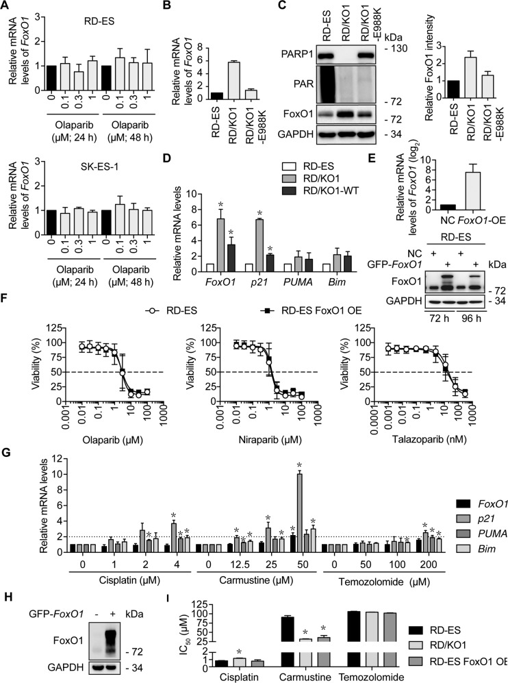
Poly(ADP-ribose) polymerase 1 (PARP1) regulates gene transcription in addition to functioning as a DNA repair factor. Forkhead box O1 (FoxO1) is a transcription factor involved in extensive biological processes. Here, we report that PARP1 binds to two separate motifs on the FoxO1 promoter and represses its transcription in a polymerase-independent manner. Using PARP1-knock out (KO) cells, wild-type-PARP1-complemented cells and catalytic mutant PARP1E988K-reconstituted cells, we investigated transcriptional regulation by PARP1. PARP1 loss led to reduced DNA damage response and ~362-fold resistance to five PARP inhibitors (PARPis) in Ewing sarcoma cells. RNA sequencing showed 492 differentially expressed genes in a PARP1-KO subline, in which the FoxO1 mRNA levels increased up to more than five times. The change in the FoxO1 expression was confirmed at both mRNA and protein levels in different PARP1-KO and complemented cells. Moreover, exogenous PARP1 overexpression reduced the endogenous FoxO1 protein in RD-ES cells. Competitive EMSA and ChIP assays revealed that PARP1 specifically bound to the FoxO1 promoter. DNase I footprinting, mutation analyses, and DNA pulldown FREP assays showed that PARP1 bound to two particular nucleotide sequences separately located at -813 to -826 bp and -1805 to -1828 bp regions on the FoxO1 promoter. Either the PARPi olaparib or the PARP1 catalytic mutation (E988K) did not impair the repression of PARP1 on the FoxO1 expression. Exogenous FoxO1 overexpression did not impair cellular PARPi sensitivity. These findings demonstrate a new PARP1-gene promoter binding mode and a new transcriptional FoxO1 gene repressor.


Conflict of interest statement

The authors declare that they have no conflict of interest.

Figures

Fig. 1
Fig. 1. Characterization of PARP1-knockout (KO) (PARP1/KO) variants.
a Levels of PARP1 and PAR were detected by western blotting in different PARP1/KO variants (KO1 and KO2) of RD-ES and SK-ES-1 cells exposed to 200 μM H2O2 for 5 min. b Accumulation of γH2AX was reduced in PARP1/KO cells relative to parental cells treated with olaparib (0, 1, 3, or 10 μM). c Levels of DNA repair-related proteins in the RD-ES, RD/KO1, Cri/KO, and RD/KO1-WT cells were determined by western blotting. d Changes in PARPi sensitivity in response to PARP1 loss and PARP1 reconstitution. IC50 values from three independent experiments were expressed as mean ± SD. Error bars represent the SD. The resistance factor (RF) is the ratio of the averaged IC50 value of indicated PARPi in given cells to that of the same PARPi in RD-ES cells. e Volcano plots of the differentially expressed genes in RD/KO1 cells [log2 fold change >1 with statistical significance (p < 0.05)] detected by RNA-seq. Significantly upregulated and downregulated genes were colored in red and blue, respectively. X axis: log2 fold change of gene expression. Y axis: statistical significance of the differential expression in the scale of −log10 (p value). f Hierarchical clustered heatmap of differentially expressed genes in PARP1 loss cells: rows represent cell lines and columns represent genes. Genes with similar expression patterns are within the same cluster and close to each other, and they may have similar functions or participate in the same biological processes. In clustering analysis, high expression and low expression genes are colored in red and blue, respectively (Genes were shown in Supplementary Table S1 from top to bottom). g Differentially expressed genes in RD/KO1 cells involved in “pathways in cancer” and “regulation of sequence-specific DNA binding transcription factor activity” were plotted in a Venn diagram to display commonly affected genes (Genes were shown in Supplementary Table S2).
Fig. 2
Fig. 2. PARP1 loss increases FoxO1 expression.
a Confirmation of some results from RNA-seq by RT-qPCR in different cells. Log2 mRNA levels of TNFAIP3, IκBα, and NF-κB1 in PARP1-KO or complemented cells were normalized to that in corresponding parental cells. Error bars represent the SD. b Confirmation of some results from RNA-seq by western blotting in indicated cells. c Loss of PARP1 increased mRNA and protein levels of FoxO1, which was prevented, at least partially, by PARP1 complementation. The mRNA levels of FoxO1 were detected by RT-qPCR and normalized to that in the corresponding parental cells; Error bars represent the SD. *, p < 0.05. Protein levels of FoxO1 were detected by western blotting. d After RD-ES cells were transfected with GFP-PARP1 cDNA for 72 h, protein levels of FoxO1 were determined by western blotting. The relative FoxO1 levels were presented as the ratio of (FoxO1/GAPDH)GFP-PARP1/(FoxO1/GAPDH)CON when the value of (FoxO1/GAPDH)CON was normalized as 1.
Fig. 3
Fig. 3. PARP1 binding to particular regions on the FoxO1 promoter.
a Schematic representation of the locations of particular nucleotide fragments (FoxO1-L, -M, and -R) on the FoxO1 promoter analyzed by EMSA. b The binding of purified PARP1 to particular nucleotide fragments (FoxO1-L, -M, and -R) were analyzed by EMSA. For the competition assays, 40-fold excess of unlabeled DNA fragments were added to the reaction mixture before adding FAM-labeled probes, and the labeled PARP1-FoxO1 complexes were almost completely displaced. c RD-ES, RD/KO1, and RD/KO1-WT cells were subjected to ChIP analyses using the antibody against PARP1 and an isotype-matched IgG as a negative control. The association of PARP1 with the FoxO1 gene promoter was quantified by RT-qPCR using primers targeting FoxO1-L, -M, and -R, respectively. Error bars represent the SD. d Identification of PARP1-protected regions on FoxO1-L and FoxO1-R by DNase I footprinting assays. Electropherograms showed the whole region of the FoxO1-L/R after digestion with DNase I following incubation in the presence (blue) or absence (red) of PARP1. The DNA sequences of the PARP1-protected regions were marked with dashed rectangles and denoted as FoxO1-L-B (−813TCACTGTATTCTT−826) and FoxO1-R-B (−1805TCTTGTGGTCTCTTCACGTTTAC−1828).
Fig. 4
Fig. 4. PARP1 binding to the specific DNA sequences on the FoxO1 promoter.
a The FAM-labeled probes containing sequences of FoxO1-L-B (left) and FoxO1-R-B (right) and corresponding mutated probes (underlined; FoxO1-L-B-M and FoxO1-R-B-M), and the deleted sequences (line-through; FoxO1-L-B-D and FoxO1-R-B-D). b EMSA was carried out using normal or mutated FAM-labeled probes. The amount of DNA-protein complexes detected in FAM-labeled mutant probes was similar to that in FAM-labeled normal probes. c EMSA was carried out using FAM-labeled normal and deletion probes. Fewer DNA-protein complexes were detected with FAM-labeled deletion probes than the FAM-labeled normal probes followed by PARP1 incubation. d Schematic representation of the flanking restriction enhanced pulldown (FREP). A biotinylated DNA fragment is conjugated to streptavidin-coated magnetic Dynabeads (Invitrogen, Carlsbad, CA). This fragment is engineered to include the FoxO1-L-B or FoxO1-R-B specific (“bait”) sequence (black dashed box), flanked by restriction enzyme cleavage sites for BamH I proximally (gray dashed box) and EcoR I distally (gray box). DNA-beads are mixed with PARP1 protein. A free non-biotinylated FoxO1-L-B or FoxO1-R-B DNA fragment can be included in the control reaction at this stage as a specific competitor. Magnetic separation and wash remove non-DNA binding PARP1 protein. EcoR I digestion releases 3′ DNA end-binding PARP1, and BamH I digestion separates the sequence-specific FoxO1-L-B or FoxO1-R-B binding PARP1 from the 5′ DNA and Dynabeads. Western blotting identifies PARP1 binding to FoxO1-L-B or FoxO1-R-B. e The PARP1-DNA complexes cut with EcoR I and BamH I were detected by western blotting. The relative levels of FoxO1-bound PARP1 were presented as the ratio of FoxO1-bound PARP1 band intensity/PARP1 input band intensity when the value of PARP1 input band intensity was normalized as l. Lane 1: PARP1 inputs, lane 2: labeled FoxO1-L-B-beads, lane 3: PARP1 with labeled FoxO1-L-B-beads, lane 4: PARP1 with labeled FoxO1-L-B-beads and cold competitor (40-fold excess of free FoxO1-L-B-beads), lane 5: labeled FoxO1-R-B-beads, lane 6: PARP1 with labeled FoxO1-R-B-beads, lane 7: PARP1 with labeled FoxO1-R-B-beads and cold competitor (40-fold excess of free FoxO1-R-B-beads), lane 8: a labeled non-specific DNA sequence (NS-beads) and lane 9: PARP1 with labeled NS-beads.
Fig. 5
Fig. 5. The expression of FoxO1 regulated by PARP1 is independent of its catalytic activity and FoxO1 does not affect the sensitivity of RD-ES cells to PARP inhibitors.
a RD-ES (upper) and SK-ES-1 (lower) cells were incubated in the indicated concentrations of olaparib for 24 h or 48 h. Then, mRNA levels of FoxO1 were detected by RT-qPCR. b mRNA levels of FoxO1 in RD/KO1 cells and their stably-transfected with mutated-PARP1 cDNA (E988K) variants were detected by RT-qPCR. c Protein levels of FoxO1 in RD/KO1 cells and their stably-transfected with mutated-PARP1 cDNA (E988K) variants were detected by western blotting. The relative FoxO1 levels were presented as the ratio of (FoxO1/GAPDH)KO1 or E988K/(FoxO1/GAPDH)RD-ES when the value of (FoxO1/GAPDH)RD-ES was normalized as l. Data were expressed as mean ± SD from three independent experiments. d Effects of PARP1 loss and PARP1 reconstitution on the expression of FoxO1 target genes. mRNA levels were detected by RT-qPCR. *, p < 0.05. e RD-ES cells were transfected with GFP-FoxO1 cDNA for 72 h and 96 h, and the mRNA (left) and protein (right) levels of FoxO1 were detected by RT-qPCR and western blotting, respectively. f Survival curves of olaparib, niraparib and talazoparib-treated RD-ES and FoxO1-overexpressed RD-ES (RD-ES FoxO1 OE) cells assessed by CCK-8 assays. Error bars represent the SD. g Changes of the mRNA levels of FoxO1 and its target genes. RD-ES cells were treated with cisplatin, carmustine and temozolomide for 12 h. Then, mRNA levels of FoxO1, p21, PUMA and Bim were detected by RT-qPCR and normalized to those in RD-ES cells without any treatments. *, p < 0.05. h FoxO1 overexpression by transfecting GFP-FoxO1 into RD-ES cells was determined by western blotting. i Sensitivity of RD-ES, RD/KO1 and RD-ES FoxO1 OE cells to cisplatin, carmustine and temozolomide. Cells were exposed to gradient concentrations of the tested agents for 72 h. IC50 values from three independent experiments were expressed as mean ± SD. Error bars represent the SD. *, p < 0.05.

References

    1. Ding J, Miao ZH, Meng LH, Geng MY. Emerging cancer therapeutic opportunities target DNA-repair systems. Trends Pharmacol. Sci. 2006;27:338–344. doi: 10.1016/j.tips.2006.04.007. - DOI - PubMed
    1. He JX, Yang CH, Miao ZH. Poly(ADP-ribose) polymerase inhibitors as promising cancer therapeutics. Acta Pharmacol. Sin. 2010;31:1172–1180. doi: 10.1038/aps.2010.103. - DOI - PMC - PubMed
    1. Wang YQ, et al. An update on poly(ADP-ribose)polymerase-1 (PARP-1) inhibitors: opportunities and challenges in cancer therapy. J. Med. Chem. 2016;59:9575–9598. doi: 10.1021/acs.jmedchem.6b00055. - DOI - PubMed
    1. Chen HD, et al. Increased PARP1-DNA binding due to autoPARylation inhibition of PARP1 on DNA rather than PARP1-DNA trapping is correlated with PARP1 inhibitor’s cytotoxicity. Int. J. Cancer. 2019;145:714–727. doi: 10.1002/ijc.32131. - DOI - PubMed
    1. Yuan B, et al. Poly(ADP-ribose)polymerase (PARP) inhibition and anticancer activity of simmiparib, a new inhibitor undergoing clinical trials. Cancer Lett. 2017;386:47–56. doi: 10.1016/j.canlet.2016.11.010. - DOI - PubMed

MeSH terms

Substances