. 2020 Jan 28;10(1):1311.
doi: 10.1038/s41598-020-58180-7.

Large deletions in immunoglobulin genes are associated with a sustained absence of DNA Polymerase η

Affiliations

Large deletions in immunoglobulin genes are associated with a sustained absence of DNA Polymerase η

Leticia K Lerner et al. Sci Rep. .

Abstract

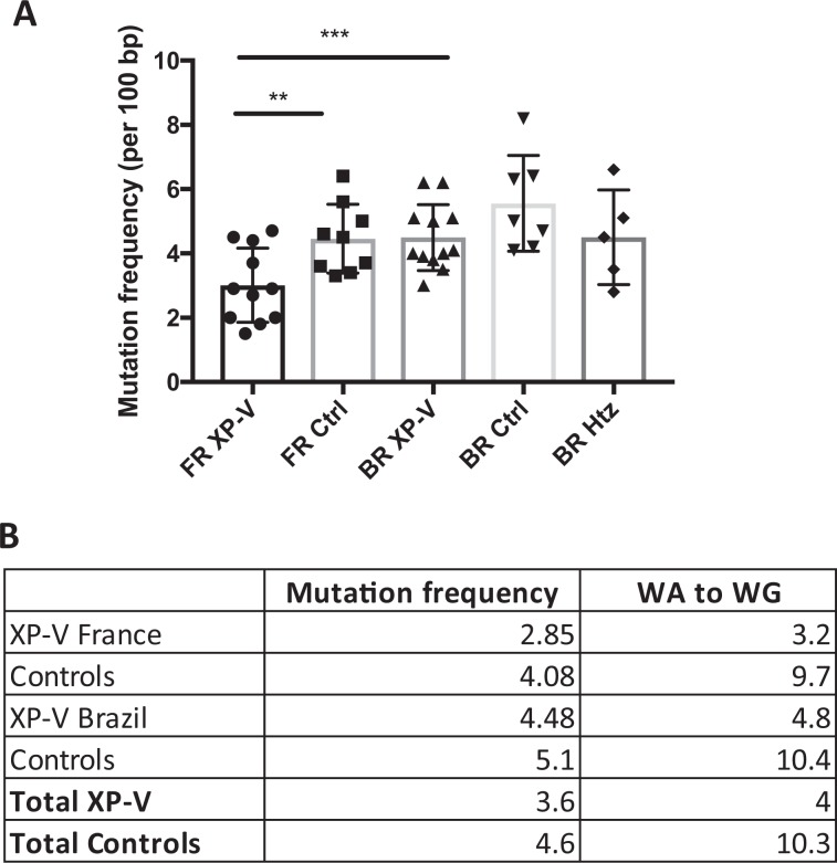
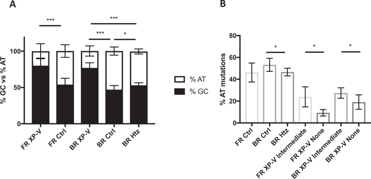
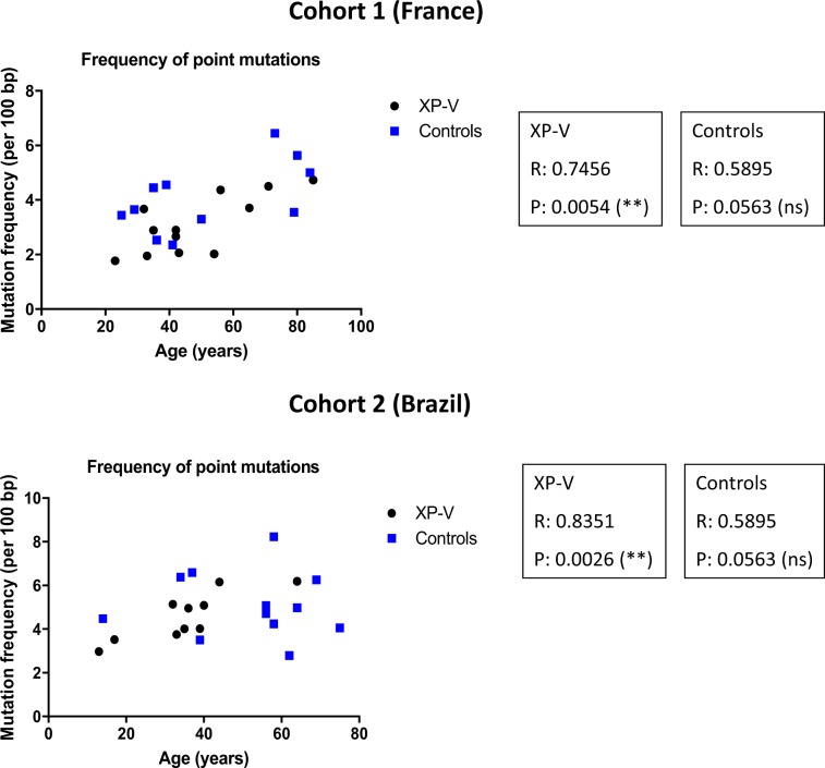
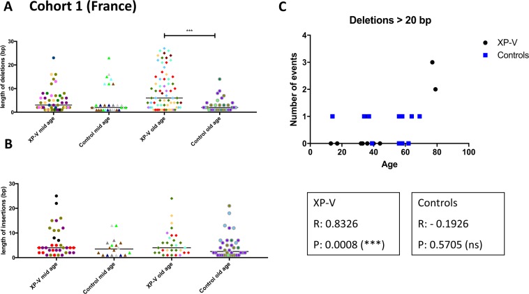
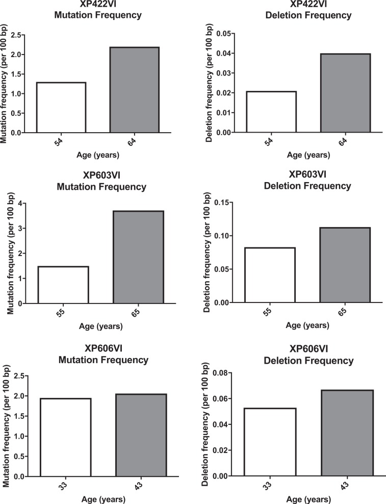
Somatic hypermutation of immunoglobulin genes is a highly mutagenic process that is B cell-specific and occurs during antigen-driven responses leading to antigen specificity and antibody affinity maturation. Mutations at the Ig locus are initiated by Activation-Induced cytidine Deaminase and are equally distributed at G/C and A/T bases. This requires the establishment of error-prone repair pathways involving the activity of several low fidelity DNA polymerases. In the physiological context, the G/C base pair mutations involve multiple error-prone DNA polymerases, while the generation of mutations at A/T base pairs depends exclusively on the activity of DNA polymerase η. Using two large cohorts of individuals with xeroderma pigmentosum variant (XP-V), we report that the pattern of mutations at Ig genes becomes highly enriched with large deletions. This observation is more striking for patients older than 50 years. We propose that the absence of Pol η allows the recruitment of other DNA polymerases that profoundly affect the Ig genomic landscape.


Conflict of interest statement

The authors declare no competing interests.

Figures

Figure 1
Figure 1
Mutation frequency is lower in XP-V patients than in controls. (A) Mutation frequency (per 100 bp) of XP-V patients (FR XP-V) and controls (FR Ctrl) of the French cohort, as well as XP-V patients (BR XP-V), wild-type (POLH homozygous) (BR Ctrl) and carrier (POLH heterozygous) (BR Htz) controls from the Brazilian cohort. **p = 0.01 and ***p = 0.0037 (Unpaired T-test). (B) SHM mutation frequency (per 100 bp), percentages of transitions and transversions and mutations in WA to WG motifs of XP-V patients, carriers and the corresponding controls.
Figure 2
Figure 2
Percentage of AT mutations is proportional to DNA polymerase η activity. (A) SHM profile as percentage of AT: GC mutations of XP-V patients (FR XP-V) and controls (FR Ctrl) of the French cohort, as well as XP-V patients (BR XP-V), wild-type (POLH homozygous)(BR Ctrl) and carrier (POLH heterozygous)(BR Htz) controls from the Brazilian cohort. *p<0.05  and ***p<0.0001 (Unpaired T-test). (B) Percentage of AT mutations of POLH WT controls from the French (FR Ctrl) and Brazilian (BR Ctrl) cohorts, POLH heterozygous (carrier) controls from the Brazilian cohort (BR Htz), XP-V patients with an intermediate pol η activity of the French (FR XP-V Intermediate) and Brazilian (BR XP-V Intermediate) cohort, and XP-V patients with no detectable pol η activity from the French (FR XP-V None) and Brazilian (BR XP-V None) cohorts. *p = 0.0418 (Unpaired T-test).
Figure 3
Figure 3
Correlation between frequency of point mutations and age in XP-V patients. Frequency of point mutation (per 100 bp) was plotted against age (years) of XP-V patients and controls of the French (upper panel) and Brazilian (bottom panel) cohorts. Values of Pearson correlation (R) and p values are indicated for each group (XP-V and controls) of both cohorts.
Figure 4
Figure 4
XP-V patients over 50 years of age present higher frequency of large deletions (larger than 10 bp) compared to age-matched controls. (A) Size of deletions (in base pairs) of XP-V patients (older than 50 years) and controls of the French cohort. Mid age: younger than 50 years. Old age: older than 50 years. ***p < or = 0.0005 (1-way Anova test). Each color represents one patient/control. (B) Size of insertions of XP-V patients (older than 50 years) and controls of the French cohort. Each color represents one patient/control. (C) Number of large deletions (larger than 20 bp) was plotted against age (years) of XP-V patients and controls of the Brazilian cohort. Values of Pearson correlation (R) and p values are indicated for each group (XP-V and controls).
Figure 5
Figure 5
10 year follow up of 3 XP-V patients show increased mutation and deletion frequencies. Mutation and deletion frequencies (per 100 bp) of the three XP-V patients from the French cohort reanalyzed 10 years later, showing values for the first and the second samplings.

References

    1. DiGiovanna JJ, Kraemer KH. Shining a light on xeroderma pigmentosum. J. Invest. Dermatol. 2012;132:785–796. doi: 10.1038/jid.2011.426. - DOI - PMC - PubMed
    1. De Weerd-Kastelein EA, Keijzer W, Bootsma D. Genetic heterogeneity of xeroderma pigmentosum demonstrated by somatic cell hybridization. Nature. New Biol. 1972;238:80–83. doi: 10.1038/newbio238080a0. - DOI - PubMed
    1. de Laat WL, Jaspers NG, Hoeijmakers JH. Molecular mechanism of nucleotide excision repair. Genes Dev. 1999;13:768–785. doi: 10.1101/gad.13.7.768. - DOI - PubMed
    1. Stary A, Sarasin A. Molecular mechanisms of UV-induced mutations as revealed by the study of DNA polymerase eta in human cells. Res. Microbiol. 2002;153:441–445. doi: 10.1016/S0923-2508(02)01343-8. - DOI - PubMed
    1. Opletalova K, et al. Correlation of phenotype/genotype in a cohort of 23 xeroderma pigmentosum-variant patients reveals 12 new disease-causing POLH mutations. Hum. Mutat. 2014;35:117–128. doi: 10.1002/humu.22462. - DOI - PubMed

Supplementary concepts