. 2020 Jan 28;10(1):1333.
doi: 10.1038/s41598-020-58068-6.

AMPA receptor deletion in developing MGE-derived hippocampal interneurons causes a redistribution of excitatory synapses and attenuates postnatal network oscillatory activity

Affiliations

AMPA receptor deletion in developing MGE-derived hippocampal interneurons causes a redistribution of excitatory synapses and attenuates postnatal network oscillatory activity

Gülcan Akgül et al. Sci Rep. .

Abstract

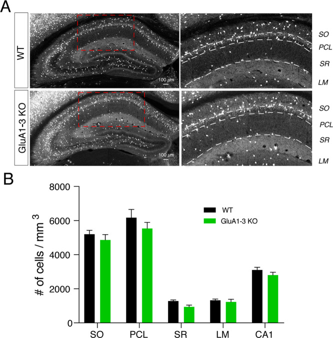
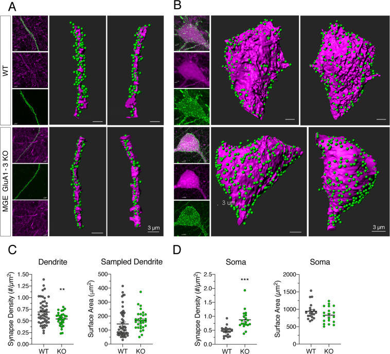
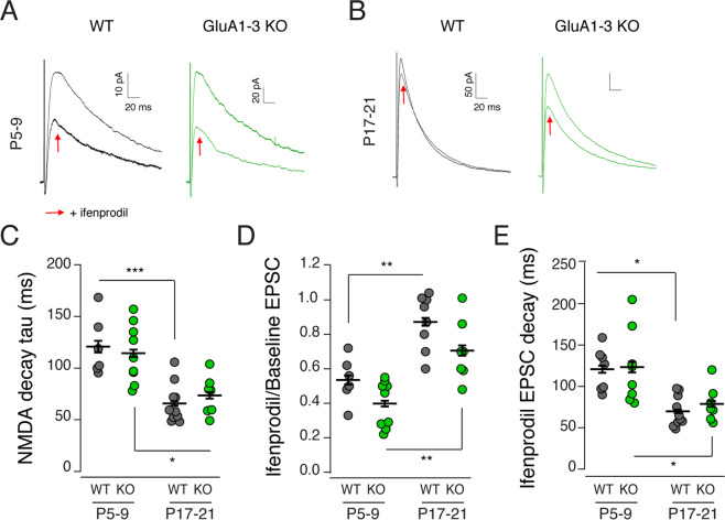
Inhibitory interneurons derived from the medial ganglionic eminence represent the largest cohort of GABAergic neurons in the hippocampus. In the CA1 hippocampus excitatory synapses onto these cells comprise GluA2-lacking, calcium-permeable AMPARs. Although synaptic transmission is not established until early in their postnatal life, AMPARs are expressed early in development, however their role is enigmatic. Using the Nkx2.1-cre mouse line we genetically deleted GluA1, GluA2, GluA3 selectively from MGE derived interneurons early in development. We observed that the number of MGE-derived interneurons was preserved in mature hippocampus despite early elimination of AMPARs, which resulted in >90% decrease in spontaneous excitatory synaptic activity. Of particular interest, excitatory synaptic sites were shifted from dendritic to somatic locations while maintaining a normal NMDAR content. The developmental switch of NMDARs from GluN2B-containing early in development to GluN2A-containing on maturation was similarly unperturbed despite the loss of AMPARs. Early network giant depolarizing potential oscillatory activity was compromised in early postnatal days as was both feedforward and feedback inhibition onto pyramidal neurons underscoring the importance of glutamatergic drive onto MGE-derived interneurons for hippocampal circuit function.


Conflict of interest statement

The authors declare no competing interests.

Figures

Figure 1
Figure 1
The number of MGE interneurons in CA1 hippocampus remains unchanged following AMPAR deletion. Low and high resolution confocal images of hippocampi from WT and KO mouse brains show that MGE interneurons are segregated to sublayers (SO, PCL, SR, SLM) of hippocampi comparably in both lines (A). (B) Bar graph shows no significant change in total number of MGE-derived interneurons in neither the entire CA1 subfield nor any particular subfield in KO. Tissue was collected from 4–6 sections from 5 WT (black) and 5 KO (green) mouse brains.
Figure 2
Figure 2
Spontaneous excitatory input onto MGE-derived interneurons is markedly decreased following GluA1-3 loss. (A) Confocal image of CA1 hippocampus in Nkx2.1:Ai14 mouse brain. Red tdTomato signal signifies MGE-derived interneurons and the green (streptavidin-Alexa 488) single neuron is a biocytin filled interneuron recovered following electrophysiological recording. (B) Representative spontaneous EPSCs (sEPSCs) recorded from MGE-derived interneurons of CA1 hippocampi of P5-9 and P17-21 WT (Nkx2.1:Ai14) or GluA1-3 KO (Nkx2.1:Ai14:Gria1-3fl/fl) mice. *Mark individual synaptic events. Recordings were done in the presence of picrotoxin (50 μM). (C) Group data are graphed for the sEPSC frequency, amplitude, decay. *P < 0.05, ***P < 0.0005.
Figure 3
Figure 3
Loss of GluA1-3 shifts subcellular synaptic localization toward somatic sites Imaris reconstructions of PSD95-egfp expressing excitatory synapses (green fluorescent puncta) on MGE-derived interneuron dendrites running through SR of CA1 (red tdTomato fluorescence) (A) and soma located in PCL and SR of CA1 (red tdTomato fluorescence) (B) of WT and GluA1-3 KO MGE interneurons. Soma and dendrites were imaged using a Zeiss Airy LSM880 and quantified and graphed (C-D). A significant shift in subcellular localization of excitatory synapses, from dendrites (C) towards soma (D), was observed in MGE KO interneurons. Synapse density: WTsoma, 0.47 ± 0.04/µm2, n = 20 soma from 2 animals; KOsoma, 0.88 ± 0.08/µm2, n = 19 soma from 2 animals, P < 0.0001; WTdendrite, 0.69 ± 0.04/µm2, n = 50 dendritic segments from 2 animals; KOdendrite, 0.54 ± 0.03/µm2, n = 30 dendritic segments from 2 animals, P = 0.0019. Soma size, WT: 945 ± 53 µm2, n = 20 soma from 2 animals; KO: 833 ± 0.03 µm2, n = 19 soma from 2 animals; P = 0.14. Dendrite surface size sampled, WT: 144 ± 14 µm2, n = 50 dendritic segments from 2 animals; KO: 170 ± 12 µm2, n = 30 dendritic segments from 2 animals; P = 0.16. Unpaired t test with Welch’s correction, two-tailed. *P < 0.05, ***P < 0.005.
Figure 4
Figure 4
Excitatory synaptic input onto MGE interneurons is critical for GDC generation. GDCs recorded from P5-10 CA3 and CA1 pyramidal neurons of WT (black), MGE GluA1-3 KO (green) and CGE GluA1-3 KO (blue). Upper traces show 120 sec of recording and lower traces show the enlarged image of a 2 sec recording focusing on the representative single event marked by a * in the respective upper trace (A). Data were plotted and analyzed in consecutive two day periods to illustrate changes in GDC frequency, amplitude and charge transfer across a 6 day developmental window (B). **P < 0.005, ***P < 0.0005.
Figure 5
Figure 5
Loss of excitatory input onto MGE interneurons results in a reduction of both feedforward and feedback GABAergic input onto CA1 pyramidal neurons. (A) Schematic depicting the loss of AMPARs at the postsynaptic site of excitatory synapses onto KO MGE interneuron (left panels) and circuit arrangement for feedforward and feedback pathways in CA1 hippocampus (right panels). Stimulating electrodes were placed in the Schaffer collateral pathway to assay disynaptic feedforward inhibition provided to CA1 pyramidal neuron, and in alveus to antidromically activate the axons of pyramidal cells which then trigger disynaptic feedback inhibition. (B) Representative Schaffer collateral mediated EPSCs (eEPSC) recorded from WT and KO MGE interneurons were evoked by paired pulse stimuli (20 Hz) and show facilitation of the synaptic current. (C) Scatter plots show the group data for eEPSC paired pulse ratio (PPR), decay time constant and NMDA/AMPA ratio. The PPR of eEPSCs was higher in KO MGE interneurons compared to WT. eEPSC decay on average was similar between WT and KO but in KO it showed a wider range of values. NMDA/AMPA EPSC ratio was larger in KO compared to WT due to AMPAR loss at synapses. (D) As a consequence of the large reduction in excitatory recruitment of MGE interneurons disynaptic feedforward inhibition onto CA1 pyramidal neurons (monitored at a holding potential of −30mV. Outward IPSCs were normalized to the inward monosynaptic EPSCs) was reduced in the MGE GluA1-3 KO group. IPSC/EPSC ratios for all recordings are shown in the scatter plot for the group data. (E) Di-synaptic feedback inhibition was also reduced in MGE GluA1-3 KO CA1 hippocampi. Evoked IPSC amplitude was normalized to the slope of the population spike recorded from PCL. Normalized IPSC is shown in the scatter plot for the group data. The wildtype control dataset for both the feedforward and feedback inhibition experiments is replotted from our earlier study since all experiments were originally performed by interleaving recordings from wildtype, MGE-KO and CGE-KO animals. *P < 0.05, **P < 0.005.
Figure 6
Figure 6
The GluN2B-to-2A developmental switch occurs independently of AMPARs. Schaffer collateral evoked NMDAR-mediated synaptic responses (Vhold =  + 50 mV) were recorded from WT (black) or GluA1-3 KO MGE (green) interneurons in CA1 hippocampi at P5-9 (A) and P17-21 (B) in the presence or absence of the GluN2B specific NMDAR antagonist ifenprodil (smaller traces indicated by red arrow) (all recordings done in the presence of picrotoxin (50uM) and DNQX (10uM)). The group data showed a significant decrease in decay tau (C) of NMDAR-mediated EPSCs between P5-9 to P17-21 in both WT and KO groups. NMDAR-mediated events became significantly less sensitive to ifenprodil from P5-9 to P17-21 (D,E). However, the loss of GluARs was without any major impact on the kinetic change or sensitivity of ifenprodil across the developmental window tested. *P < 0.05, **P < 0.005, ***P < 0.0005.

References

    1. Pelkey KA, et al. Hippocampal GABAergic Inhibitory Interneurons. Physiol. Rev. 2017;97:1619–1747. doi: 10.1152/physrev.00007.2017. - DOI - PMC - PubMed
    1. Tricoire L, et al. A blueprint for the spatiotemporal origins of mouse hippocampal interneuron diversity. J. Neurosci. 2011;31:10948–10970. doi: 10.1523/JNEUROSCI.0323-11.2011. - DOI - PMC - PubMed
    1. Matta JA, et al. Developmental origin dictates interneuron AMPA and NMDA receptor subunit composition and plasticity. Nat. Neurosci. 2013;16:1032–1041. doi: 10.1038/nn.3459. - DOI - PMC - PubMed
    1. Pelkey KA, et al. Pentraxins coordinate excitatory synapse maturation and circuit integration of parvalbumin interneurons. Neuron. 2015;85:1257–1272. doi: 10.1016/j.neuron.2015.02.020. - DOI - PMC - PubMed
    1. Akgul G, McBain CJ. Diverse roles for ionotropic glutamate receptors on inhibitory interneurons in developing and adult brain. J. Physiol. 2016;594:5471–5490. doi: 10.1113/JP271764. - DOI - PMC - PubMed

LinkOut - more resources