. 2020 Jan 28;10(1):1296.
doi: 10.1038/s41598-020-58385-w.

Prematurely aging mitochondrial DNA mutator mice display subchondral osteopenia and chondrocyte hypertrophy without further osteoarthritis features

Affiliations

Prematurely aging mitochondrial DNA mutator mice display subchondral osteopenia and chondrocyte hypertrophy without further osteoarthritis features

Jeroen Geurts et al. Sci Rep. .

Abstract

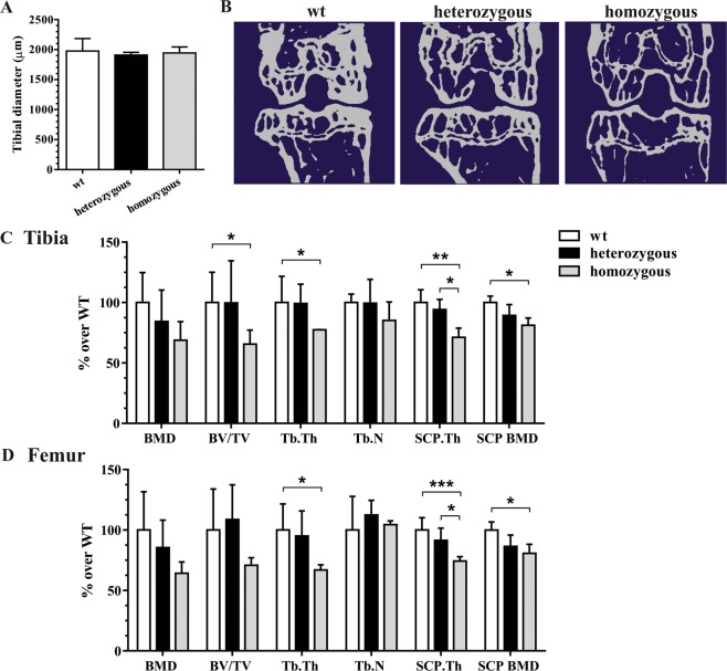
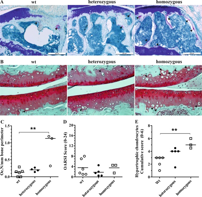
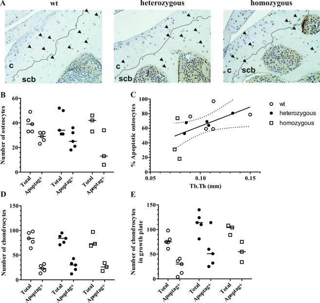
Mitochondrial mutations and dysfunction have been demonstrated in several age-related disorders including osteoarthritis, yet its relative contribution to pathogenesis remains unknown. Here we evaluated whether premature aging caused by accumulation of mitochondrial DNA mutations in PolgD275A mice predisposes to the development of knee osteoarthritis. Compared with wild type animals, homozygous PolgD275A mice displayed a specific bone phenotype characterized by osteopenia of epiphyseal trabecular bone and subchondral cortical plate. Trabecular thickness was significantly associated with osteocyte apoptosis rates and osteoclasts numbers were increased in subchondral bone tissues. While chondrocyte apoptosis rates in articular and growth plate cartilage were similar between groups, homozygous mitochondrial DNA mutator mice displayed elevated numbers of hypertrophic chondrocytes in articular calcified cartilage. Low grade cartilage degeneration, predominantly loss of proteoglycans, was present in all genotypes and the development of osteoarthritis features was not found accelerated in premature aging. Somatically acquired mitochondrial DNA mutations predispose to elevated subchondral bone turnover and hypertrophy in calcified cartilage, yet additional mechanical or metabolic stimuli would seem required for induction and accelerated progression of aging-associated osteoarthritis.


Conflict of interest statement

The authors declare no competing interests.

Figures

Figure 1
Figure 1
Measurement of tibial diameter at the transaxial reference plane 1 mm below the growth plate (A). Representative two-dimensional μCT images of the subchondral cortical plate and epiphyseal trabecular bone of the tibiofemoral joint in wild type, heterozygous and homozygous mutator mice (B). Quantitative assessment of trabecular and cortical bone parameters in tibial (C) and femoral compartments (D). *P < 0.05 and **P < 0.01 by One-way ANOVA.
Figure 2
Figure 2
Representative TRAP-stained tissue sections of the epiphyseal bones of the tibial compartment in wild type, heterozygous and homozygous mutator mice (A). Increased TRAP-staining (arrowheads) was present at the subchondral cortical plate and trabecular bone of homozygous mutants. Representative images of Safranin-O-stained tissue sections of the tibiofemoral joints. Hypertrophic chondrocytes were observed in calcified cartilage (arrowheads) (B). Histomorphometric assessment of osteoclast numbers per mm bone perimeter confirmed a significant increase of osteoclasts in homozygous mutator mice compared to wild type littermates (C). Cumulative OARSI scores of tibiofemoral joint compartments confirmed a lack of severe OA in all groups (D). Numbers of hypertrophic chondrocytes were elevated in tibiofemoral cartilage (E) **P < 0.01.
Figure 3
Figure 3
Representative tissue sections stained for apoptotic cells (arrowheads) using an indirect TUNEL method (A). Apoptotic (Apoptag+) and total amount of cells were counted in subchondral bone (B) articular cartilage (D) and growth plate cartilage (E). Correlation between femorotibial trabecular thickness measurements and percentage of apoptotic osteocytes (C). The regression line with 95% confidence intervals are displayed. c: cartilage, scb: subchondral cortical bone.

References

    1. Fernandez-Moreno M, et al. Mitochondrial DNA haplogroups influence the risk of incident knee osteoarthritis in OAI and CHECK cohorts. A meta-analysis and functional study. Annals of the rheumatic diseases. 2017;76:1114–1122. doi: 10.1136/annrheumdis-2016-210131. - DOI - PubMed
    1. Delco, M. L., Bonnevie, E. D., Bonassar, L. J. & Fortier, L. A. Mitochondrial dysfunction is an acute response of articular chondrocytes to mechanical injury. Journal of orthopaedic research: official publication of the Orthopaedic Research Society, 10.1002/jor.23651 (2017). - PMC - PubMed
    1. Ansari M.Y., Khan N.M., Ahmad I., Haqqi T.M. Parkin clearance of dysfunctional mitochondria regulates ROS levels and increases survival of human chondrocytes. Osteoarthritis and Cartilage. 2018;26(8):1087–1097. doi: 10.1016/j.joca.2017.07.020. - DOI - PMC - PubMed
    1. Blanco FJ, Rego I, Ruiz-Romero C. The role of mitochondria in osteoarthritis. Nature reviews. Rheumatology. 2011;7:161–169. doi: 10.1038/nrrheum.2010.213. - DOI - PubMed
    1. Rego-Perez I, et al. Mitochondrial DNA haplogroups associated with MRI-detected structural damage in early knee osteoarthritis. Osteoarthritis and cartilage. 2018;26:1562–1569. doi: 10.1016/j.joca.2018.06.016. - DOI - PubMed

MeSH terms