. 2020 Jan 28;3(1):46.
doi: 10.1038/s42003-020-0772-0.

Structural insights into ATP hydrolysis by the MoxR ATPase RavA and the LdcI-RavA cage-like complex

Affiliations

Structural insights into ATP hydrolysis by the MoxR ATPase RavA and the LdcI-RavA cage-like complex

Matthew Jessop et al. Commun Biol. .

Abstract

The hexameric MoxR AAA+ ATPase RavA and the decameric lysine decarboxylase LdcI form a 3.3 MDa cage, proposed to assist assembly of specific respiratory complexes in E. coli. Here, we show that inside the LdcI-RavA cage, RavA hexamers adopt an asymmetric spiral conformation in which the nucleotide-free seam is constrained to two opposite orientations. Cryo-EM reconstructions of free RavA reveal two co-existing structural states: an asymmetric spiral, and a flat C2-symmetric closed ring characterised by two nucleotide-free seams. The closed ring RavA state bears close structural similarity to the pseudo two-fold symmetric crystal structure of the AAA+ unfoldase ClpX, suggesting a common ATPase mechanism. Based on these structures, and in light of the current knowledge regarding AAA+ ATPases, we propose different scenarios for the ATP hydrolysis cycle of free RavA and the LdcI-RavA cage-like complex, and extend the comparison to other AAA+ ATPases of clade 7.


Conflict of interest statement

The authors declare no competing interests.

Figures

Fig. 1
Fig. 1. Cryo-EM structure of the LdcI–RavA cage-like complex.
a Pseudo-atomic model of the LdcI–RavA complex, based on flexible fitting of crystal structures of RavA (PDB ID: 3NBX) and LdcI (PDB ID: 3N75)12. The cryo-EM map used for fitting corresponds to the “Class 1” map after 3D classification (containing two LdcI decamers and one RavA hexamer, see panel b) to which C5 symmetry has been applied. Two LdcI decamers (coloured yellow and orange) and five spiral RavA hexamers (individually coloured light to dark blue) are shown as cartoons. Top and side views are shown, as well as a cut-away side view displaying the inner cavity of the cage. A dashed box (b) shows one RavA monomer with annotations for the different domains: AAA+ module (green), triple helical domain (red), and LARA domain (blue). The seam in the spiral RavA hexamer is indicated by a dashed line. c, d Classes 1 (c) and 2 (d) obtained after C5 symmetry expansion followed by a masked 3D classification without angular search in RELION-2.0, resulting in C1 asymmetric maps. For each class, a post-processed cryo-EM map is shown (left) along with a fit of two LdcI decamers (yellow and orange) and one RavA hexamer (Class 1: cyan, Class 2: dark blue). Dashed lines indicate the positions of the masked-out RavA hexamers during symmetry expansion. On the right, side and top views of the fits are shown, with panels (e)–(h) (dashed boxes) focusing on specific LARA domains (numbered 1–6, black circles: rigid, red circles: flexible) contacting LdcI and their corresponding fits in the EM map. Class 2 displays a 6° tilted orientation of the second LdcI decamer (coloured orange) compared to Class 1.
Fig. 2
Fig. 2. Comparison between a RavA hexamer generated from the RavA crystal structure and a fit in the cryo-EM map of the LdcI–RavA complex.
a Crystal structure of E. coli RavA (PDB ID: 3NBX), displayed as cartoons, showing the helical crystal packing of RavA crystallised in spacegroup P65. A top view along the helical screw axis of the assembly (left, with annotated unit-cell parameters) resembles a RavA hexamer. A side view of the helical assembly is shown as well (middle, with annotated unit-cell parameters), with one spiral RavA hexamer coloured dark blue. On the right, an extracted spiral hexamer (dark blue) is displayed with a slight tilt to allow visualisation of the seam, along with a schematic representation. b Comparison of a spiral RavA hexamer extracted from the crystal structure (dark blue) and a fit of RavA in the cryo-EM map of Class 1 (light blue). Side (left) and top views (right) are shown, with dashed circles around the LARA domains (numbered 1–6) to highlight the differences between the crystal structure and EM fit. The position of the seam is indicated by a dashed line. c LdcI–RavA complex obtained after fitting of structures of RavA (light blue) and LdcI (yellow and orange) in the cryo-EM map of Class 1, with LARA domains numbered as in (b).
Fig. 3
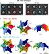
Fig. 3. Cryo-EM analysis of free RavA in the presence of nucleotide (ADP).
a, b 2D classes of untilted (a) and 30° tilted (b) datasets of free RavA in the presence of ADP. Red squares highlight classes belonging to a spiral RavA conformation, while blue squares show classes belonging to a C2-symmetric closed ring conformation of RavA. c, d 3D reconstructions of the spiral (c) and closed ring (d) RavA conformations corresponding to class 1 and 2, respectively. Individual subunits in the maps are coloured according to a rainbow colour scheme. The nucleotide-free seams in the two maps are annotated using dotted arrows.
Fig. 4
Fig. 4. Structural analysis of spiral and C2-symmetric closed ring RavA conformations.
Fit of the spiral (a) and C2-symmetric closed ring (b) conformations of RavA in their respective EM maps, displayed as cartoons. Individual RavA subunits, labelled 1–6 in the accompanying schematic representations, are coloured according to a rainbow colour scheme. Zooms show the presence or absence of ADP in the nucleotide-binding site interface formed between subunits 2–3 and 3–4 in the spiral (a) and C2-symmetric closed ring (b) conformations of RavA. The nucleotide-free seams in the two maps are annotated using black arrows. cf. Insets showing the nucleotide-binding site interface formed between subunits 1–2 (c), 4–5 (d) and 5–6 (e) of the spiral RavA conformation, and between subunits 1–2 (f) of the C2-symmetric closed ring RavA conformation.
Fig. 5
Fig. 5. Comparison between C2-symmetric closed ring conformations of RavA and ClpX.
Comparison between closed ring conformations of RavA (a) and ClpX (b), shown as cartoons with accompanying schematic representations. RavA and ClpX subunits in equivalent positions around the hexamer are given identical colours following a rainbow colour scheme. In a RavA hexamer, the active site is formed in between the large and small AAA+ domains of adjacent RavA monomers, while in ClpX the nucleotide-binding site is formed between the large and small AAA+ domains of within a single ClpX subunit. Loadable and unloadable ATP binding sites in RavA and ClpX are annotated with L and U, respectively. c, d Zooms of the nucleotide-binding interface between adjacent RavA monomers (c, coloured orange and green) and the nucleotide-binding interface within one ClpX subunit (d, coloured green). Bound ADP molecules and interacting residues of RavA or ClpX are labelled and shown as sticks. e Superposition of the large and small AAA+ domains of adjacent RavA subunits (labelled RavA and RavA) from an interface in the C2-symmetric closed ring conformation with bound ADP (“closed”conformation, coloured pink and purple) and without bound ADP (“open” conformation, coloured light and dark blue). f Superposition of the large and small AAA+ domains within one ClpX subunit containing bound ADP (“closed” conformation, coloured pink) or without bound ADP (“open” conformation, coloured light blue). Movement of the small AAA+ domains of RavA and ClpX upon nucleotide binding is shown using black arrows.
Fig. 6
Fig. 6. Characterisation of the LdcI–RavA interaction by ATPase assays and BLI binding studies.
a Effect of LdcI on the ATPase activity of RavA. ATPase activity was measured at various pH values (ranging from 5 to 9) as described in “Methods”, either for RavA alone (red) or a mix containing RavA and a three-fold molar excess of LdcI (blue). Each data point represents the average of three independent measurements. Error bars correspond to the standard deviation. The dashed inset (b) shows the percent change of RavA activity when comparing the ATPase activity of RavA alone and RavA plus a three-fold molar excess of LdcI. The statistical significance is calculated using a 2-sided T-test (2-sample unequal variance). P ≥ 0.1: not significant (ns), P ≤ 0.1: *P ≤ 0.05: **P ≤ 0.001: ***. c BLI measurements of the LdcI–RavA interaction at different pH values ranging from 5 to 8 (blue curves: experimental data, red curves: calculated fit using a 1:1 interaction model.). For each experiment, biotinylated RavA-AVITAG was immobilised on a streptavidin-coated BLI biosensor (with or without prior incubation with 1 mM ADP) followed by binding measurements using different concentrations of LdcI (500 nM, 250 nM, 125 nM, 62.5 nM and 31.25 nM). Average values for the RavA-ADP:LdcI interaction measured at four different pH values (8, 7, 6.5, 5) are: KD = 29.7 nM ± 10.0, kon = 2.68 × 10⁴ 1/Ms ± 2.13 × 10³, and kdis = 8.11 × 10⁻⁴ 1/s ± 3.41 × 10⁻⁴. The KD, kon and kdis values of the individual experiments can be found in Supplementary Table 2. d Negative-stain EM micrographs of LdcI incubated at different pH values (reproduced from Kanjee et al., with permission of the EMBO Journal). At lower pH, LdcI mainly forms decamers (pH 6.5) or stacks of decamers (pH 5), while at higher pH (pH 8) LdcI is predominantly dimeric.

Similar articles

Cited by

References

    1. Snider J, Houry WA. MoxR AAA+ ATPases: a novel family of molecular chaperones? J. Struct. Biol. 2006;156:200–209. doi: 10.1016/j.jsb.2006.02.009. - DOI - PubMed
    1. Wong KS, Houry WA. Novel structural and functional insights into the MoxR family of AAA+ ATPases. J. Struct. Biol. 2012;179:211–221. doi: 10.1016/j.jsb.2012.03.010. - DOI - PubMed
    1. Kahle M, ter Beek J, Hosler JP, Ädelroth P. The insertion of the non-heme FeBcofactor into nitric oxide reductase from P. denitrificans depends on NorQ and NorD accessory proteins. Biochim. Biophys. Acta—Bioenerg. 2018;1859:1051–1058. doi: 10.1016/j.bbabio.2018.05.020. - DOI - PubMed
    1. Tsai YCC, Lapina MC, Bhushan S, Mueller-Cajar O. Identification and characterization of multiple rubisco activases in chemoautotrophic bacteria. Nat. Commun. 2015;6:8883. doi: 10.1038/ncomms9883. - DOI - PMC - PubMed
    1. Sutter, M. et al. Structural characterization of a newly identified component of α-carboxysomes: the AAA+ domain protein CsoCbbQ. Sci. Rep. 5, 16243 (2015). - PMC - PubMed

MeSH terms