Review
. 2020 Jan 14:10:861.
doi: 10.3389/fendo.2019.00861. eCollection 2019.

Aging and Imaging Assessment of Body Composition: From Fat to Facts

Affiliations
Review

Aging and Imaging Assessment of Body Composition: From Fat to Facts

Federico Ponti et al. Front Endocrinol (Lausanne). .

Abstract

The aging process is characterized by the chronic inflammatory status called "inflammaging", which shares major molecular and cellular features with the metabolism-induced inflammation called "metaflammation." Metaflammation is mainly driven by overnutrition and nutrient excess, but other contributing factors are metabolic modifications related to the specific body composition (BC) changes occurring with age. The aging process is indeed characterized by an increase in body total fat mass and a concomitant decrease in lean mass and bone density, that are independent from general and physiological fluctuations in weight and body mass index (BMI). Body adiposity is also re-distributed with age, resulting in a general increase in trunk fat (mainly abdominal fat) and a reduction in appendicular fat (mainly subcutaneous fat). Moreover, the accumulation of fat infiltration in organs such as liver and muscles also increases in elderly, while subcutaneous fat mass tends to decrease. These specific variations in BC are considered risk factors for the major age-related diseases, such as cardiovascular diseases, type 2 diabetes, sarcopenia and osteoporosis, and can predispose to disabilities. Thus, the maintenance of a balance rate of fat, muscle and bone is crucial to preserve metabolic homeostasis and a health status, positively contributing to a successful aging. For this reason, a detailed assessment of BC in elderly is critical and could be an additional preventive personalized strategy for age-related diseases. Despite BMI and other clinical measures, such as waist circumference measurement, waist-hip ratio, underwater weighing and bioelectrical impedance, are widely used as a surrogate measure for body adiposity, they barely reflect the distribution of body fat. Because of the great advantages offered by imaging tools in research and clinics, the attention of clinicians is now moving to powerful imaging techniques such as computed tomography, magnetic resonance imaging, dual-energy X-ray absorptiometry and ultrasound to obtain a more accurate estimation of BC. The aim of this review is to present the state of the art of the imaging techniques that are currently available to measure BC and that can be applied to the study of BC changes in the elderly, outlining advantages and disadvantages of each technique.

Keywords: age-related diseases; aging; body composition; fat and lean mass; imaging techniques.


Figures

Figure 1
Figure 1
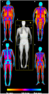
Dual-energy X-Ray absorptiometry (DXA) examination of body composition. In the center of the picture (E) is represented the skeletal map of whole body scan by DXA highlighting the standard ROIs specifics for body composition assessment (head—H, trunk—T, upper limbs—U, lower limbs—L), with the two regions at “high metabolic significance” representing by gynoid (G) and android (A) regions. On the side are depicted the soft tissue maps of whole body DXA scan (from fat mass—yellow—to bone mass—blue); in particular on the left are visualized old (A) and young (C) males (respectively upper and lower), while in the right old (B) and young (D) females (respectively upper and lower), highlighting the increase of fat mass in aging. Images are kindly provided by IRCCS Rizzoli Orthopedic Institute, Unit of Diagnostic and Interventional Radiology (2019).
Figure 2
Figure 2
CT images of the android region. CT image slices of the android region showing changes in adiposity distribution (visceral fat—arrowheads; subcutaneous fat—arrows) depending on age and sex: (A) young male, (B) old male, (C) young female, (D) old female. With advancing age, there is a redistribution of fat mass compartment with increase of visceral compartment for both sexes (in particular for males); it is also noteworthy that the subcutaneous compartment is prevalent in females, both in young and old age. Images are kindly provided by IRCCS Rizzoli Orthopedic Institute, Unit of Diagnostic and Interventional Radiology (2019).
Figure 3
Figure 3
MR T1-weighted image slices of the gynoid region showing age-related muscle changes in both sexes (poor muscle quality and fat infiltration—arrows). In addition larger subcutaneous adipose tissue are observed in the gynoid region of an old female (D) compared to a young female (C); on the contrary the representation of subcutaneous compartment in the same region is the same both for a young male (A) and an old male (B). Images are kindly provided by IRCCS Rizzoli Orthopedic Institute, Unit of Diagnostic and Interventional Radiology (2019).
Figure 4
Figure 4
Graphical summary of the standards for DXA, CT, MRI and US for the detection of metabolic dysfunction in elderly. Standards for sarcopenia, obesity and sarcopenic obesity are summarized.
Figure 5
Figure 5
The changes that usually occur with age such as overall increase of body fat and ectopic fat infiltration and the decrease of skeletal muscle should be accurately measured in order to add this information to a personalized preventive strategy to counteract age-related disease and disabilities. Although anthropometric measures, underwater weighting and electric bioimpedance represent cheap, easy and completely safe methods, they do not guarantee a high precision and accuracy to define BC compartments with none or a scarce definition. On the other hand, imaging techniques can guarantee a very high definition of body compartments either in fat or lean mass with a high accuracy and precision. However, all the imaging methods expose the subjects to low or medium levels of radiation, are not easily available and are quite expensive. Depending on the information sought, all these aspects should be taken into account when selecting the method to measure BC.

References

    1. World Health Organization World Report on Ageing and Health. World Health Organization (2015).
    1. Kennedy BK, Berger SL, Brunet A, Campisi J, Maria A, Epel ES, et al. Aging: a common driver of chronic diseases and a target for novel interventions. Cell. (2014) 159:709–13. 10.1016/j.cell.2014.10.039 - DOI - PMC - PubMed
    1. Franceschi C, Garagnani P, Parini P, Giuliani C, Santoro A. Inflammaging: a new immune–metabolic viewpoint for age-related diseases. Nat Rev Endocrinol. (2018) 14:576–90. 10.1038/s41574-018-0059-4 - DOI - PubMed
    1. Franceschi C, Bonafè M, Valensin S, Olivieri F, De Luca M, Ottaviani E, et al. . Inflamm-aging. An evolutionary perspective on immunosenescence. Ann N Y Acad Sci. (2000) 908:244–54. 10.1111/j.1749-6632.2000.tb06651.x - DOI - PubMed
    1. Franceschi C, Zaikin A, Gordleeva S, Ivanchenko M, Bonifazi F, Storci G, et al. Seminars in immunology. Semin Immunol. (2018) 40:1–5. 10.1016/j.smim.2018.10.008 - DOI - PubMed

LinkOut - more resources