

The Fungal Cell Wall: Candida, Cryptococcus, and Aspergillus Species
- PMID: 31993032
- PMCID: PMC6962315
- DOI: 10.3389/fmicb.2019.02993
The Fungal Cell Wall: Candida, Cryptococcus, and Aspergillus Species
Abstract
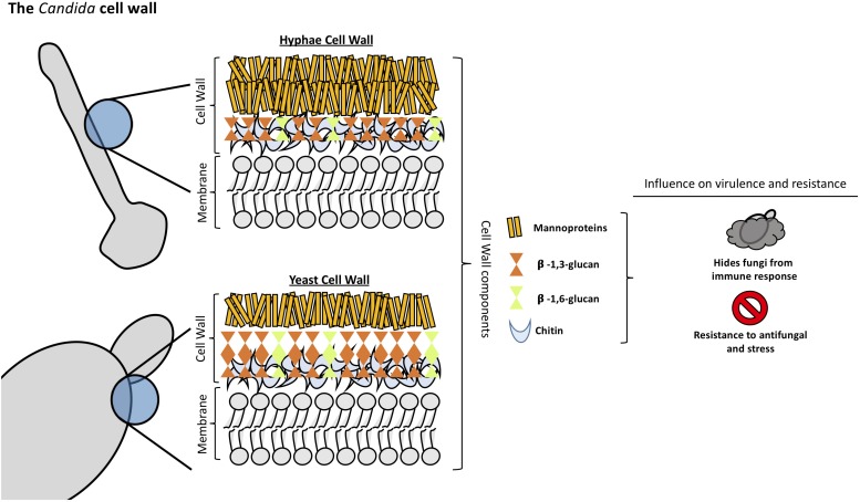
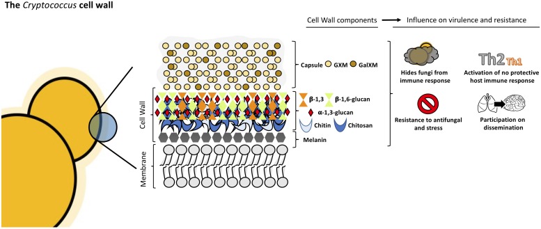
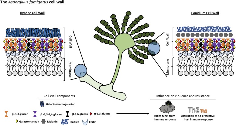
The fungal cell wall is located outside the plasma membrane and is the cell compartment that mediates all the relationships of the cell with the environment. It protects the contents of the cell, gives rigidity and defines the cellular structure. The cell wall is a skeleton with high plasticity that protects the cell from different stresses, among which osmotic changes stand out. The cell wall allows interaction with the external environment since some of its proteins are adhesins and receptors. Since, some components have a high immunogenic capacity, certain wall components can drive the host's immune response to promote fungus growth and dissemination. The cell wall is a characteristic structure of fungi and is composed mainly of glucans, chitin and glycoproteins. As the components of the fungal cell wall are not present in humans, this structure is an excellent target for antifungal therapy. In this article, we review recent data on the composition and synthesis, influence of the components of the cell wall in fungi-host interaction and the role as a target for the next generation of antifungal drugs in yeasts (Candida and Cryptococcus) and filamentous fungi (Aspergillus).
Keywords: Aspergillus; Candida; Cryptococcus; cell wall; composition; synthesis.
Copyright © 2020 Garcia-Rubio, de Oliveira, Rivera and Trevijano-Contador.
Figures
References
Publication types
LinkOut - more resources
Full Text Sources
Other Literature Sources
Research Materials