. 2019 Oct-Dec;11(4):22-32.
doi: 10.32607/20758251-2019-11-4-22-32.

Non-neutralizing Antibodies Directed at Conservative Influenza Antigens

Affiliations

Non-neutralizing Antibodies Directed at Conservative Influenza Antigens

E S Sedova et al. Acta Naturae. 2019 Oct-Dec.

Abstract

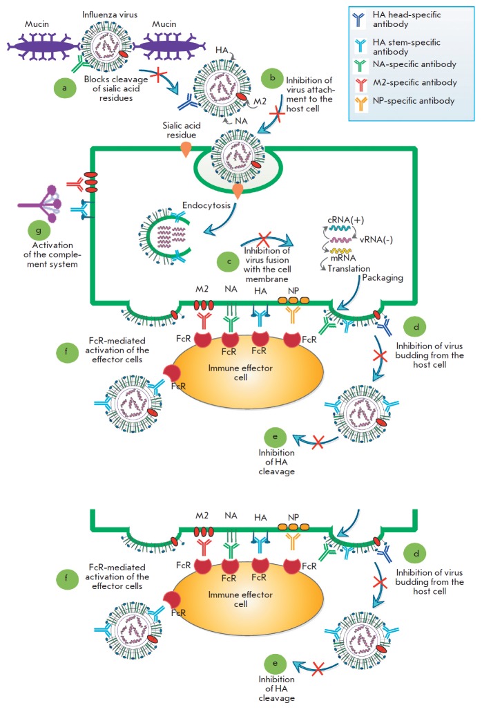
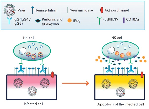
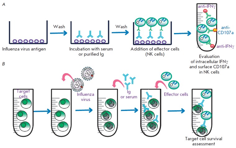
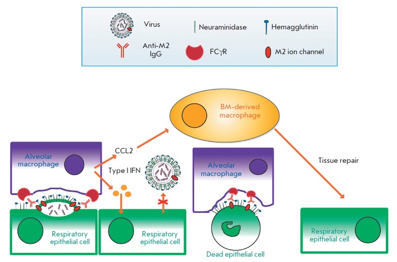
At the moment, developing new broad-spectrum influenza vaccines which would help avoid annual changes in a vaccine's strain set is urgency. In addition, developing new vaccines based on highly conserved influenza virus proteins could allow us to better prepare for potential pandemics and significantly reduce the damage they cause. Evaluation of the humoral response to vaccine administration is a key aspect of the characterization of the effectiveness of influenza vaccines. In the development of new broad-spectrum influenza vaccines, it is important to study the mechanisms of action of various antibodies, including non-neutralizing ones, as well as to be in the possession of methods for quantifying these antibodies after immunization with new vaccines against influenza. In this review, we focused on the mechanisms of anti-influenza action of non-neutralizing antibodies, such as antibody-dependent cellular cytotoxicity (ADCC), antibody-dependent cellular phagocytosis (ADCP), and antibody-mediated complement-dependent cytotoxicity (CDC). The influenza virus antigens that trigger these reactions are hemagglutinin (HA) and neuraminidase (NA), as well as highly conserved antigens, such as M2 (ion channel), M1 (matrix protein), and NP (nucleoprotein). In addition, the mechanisms of action and methods for detecting antibodies to neuraminidase (NA) and to the stem domain of hemagglutinin (HA) of the influenza virus are considered.

Keywords: antibody-dependent cellular cytotoxicity; antibody-dependent cellular phagocytosis; antibody-mediated complement-dependent cytotoxicity; broad-spectrum influenza vaccine; influenza virus.


Figures

Fig. 1
Fig. 1
Mechanisms of action of anti-influenza antibodies. The influenza virus enters the body through respiratory tract mucosa, where viral hemagglutinin (HA) binds to the terminal sialic acids of mucin. Neuraminidase (NA) releases the virus by cleaving the terminal sialic acid residues. Antibodies to neuraminidase can inhibit the reaction, and the virus would not be able to penetrate the mucous layer (a). After penetrating the mucous layer, the influenza virus binds to the sialic acids on the surface of the target cells and enters the cell by endocytosis. Neutralizing antibodies bind to influenza HA and block this process (b).The endosomes of the target cells become acidified, thus triggering the fusion of the endosomal and viral membranes via HA, which results in the release of the viral genome into the cell cytoplasm. Antibodies to the stem domain of HA can inhibit this process (c).After the synthesis of viral proteins, the internal proteins are packed into viral particles containing HA, NA, and the M2 ion channel molecules on the virion surface. On the cell surface, the HA, NA, and M2 proteins can be bound by antibodies that block the budding of viral particles. Maturing viral particles are covered by the host cell membrane as a result of the interaction between HA and sialic acids. Meanwhile, NA cleaves terminal sialic acids from the virus, while antibodies to NA can inhibit this process (d). Finally, in the matured viral particles, HA0 is cleaved into the HA1 and HA2 subunits by the host proteases that are present in the respiratory tract. Antibodies directed to the HA stem domain can block this process (e).In addition, viral antigens exposed to the surface of an infected cell (including the internal protein NP, which is detected on the surface of the infected cell) are targets for antibodies that activate effector cells via the Fc-FcR interaction (f).Antibodies directed to the viral antigens exposed on the cell surface can also activate the complement system (g)
Fig. 2
Fig. 2
The mechanism of antibody-dependent cellular cytotoxicity (ADCC) in a cell infected with an influenza virus. IgG binds to the viral antigens on the surface of the infected cell. NK cells recognize the infected cells via Fc-FcR interactions and then release cytotoxic granules and secrete antiviral cytokines
Fig. 3
Fig. 3
Evaluation of ADCC in the laboratory. (a) Evaluation of ADCC using immobilized influenza virus antigens. Firstly, antigens are immobilized, washed, and incubated with serum or IgG isolated from the blood. Secondly, unbound IgGs are washed off and effector cells (peripheral blood monocytes or isolated NK cells) are added to the antigen-antibody complex. Thirdly, after incubation, activation of the effector cells is analyzed. Activation is evaluated by adding labeled antibodies to the surface and secreted marker proteins (as a rule, the surface membrane activation marker CD107a and interferon gamma are used). (b) Evaluation of ADCC using influenza virus-infected cells or target cells expressing the major viral antigens. Cells expressing viral antigens are incubated with serum or preliminarily purified IgG. Next, effector cells are added to the antibody-treated cells and ADCC is evaluated by counting dead target antibody-treated cells
Fig. 4
Fig. 4
The mechanism of the protective action of the antibodies binding to the ectodomain of the influenza M2 ion channel. Respiratory tract epithelial cells infected with the influenza A virus expose the HA, NA, and M2 viral proteins on their surface. New viral particles are budding from the infected cells. On the surface of the budding virion, antibodies bind to the ectodomain of the M2 protein and opsonize the viral particle. These antibodies activate alveolar macrophages via Fcγ receptors. Activated macrophages are able to phagocytize budding virions and fragments of the cell membrane containing the M2 protein. Dying infected cells can be also opsonized by antibodies to the M2 ectodomain and phagocytosed by alveolar macrophages via the FcγR-dependent pathway. Activated macrophages also produce type I interferons, which possess antiviral activity and regulate the expression of chemokine CCL2, which attracts bone marrow macrophages promoting tissue repair

References

    1. Paules C., Subbarao K.. Lancet. 2017;390:697–708. - PubMed
    1. Sedova E.S., Shcherbinin D.N., Migunov A.I., Smirnov Iu.A., Logunov D.Iu., Shmarov M.M., Tsybalova L.M., Naroditskiî B.S., Kiselev O.I., Gintsburg A.L.. Acta Naturae. 2012;4(4):17–27. - PMC - PubMed
    1. Krammer F.. Nat. Rev. Immunol. 2019;19(6):383–397. - PubMed
    1. Shcherbinin D.N., Alekseeva S.V., Shmarov M.M., Smirnov Y.A., Naroditskiy B.S., Gintsburg A.L.. Acta Naturae. 2016;8(1):13–20. - PMC - PubMed
    1. Truelove S., Zhu H., Lessler J., Riley S., Read J.M., Wang S., Kwok K.O., Guan Y., Jiang C.Q., Cummings D.A., Influenza Other Respi. Viruses. 2016;10:518–524. - PMC - PubMed

LinkOut - more resources