Review
. 2020 Mar 1;9(3):127-143.
doi: 10.1089/wound.2019.1021. Epub 2020 Jan 24.

Skin Structure-Function Relationships and the Wound Healing Response to Intrinsic Aging

Affiliations
Review

Skin Structure-Function Relationships and the Wound Healing Response to Intrinsic Aging

Michael J Blair et al. Adv Wound Care (New Rochelle). .

Abstract

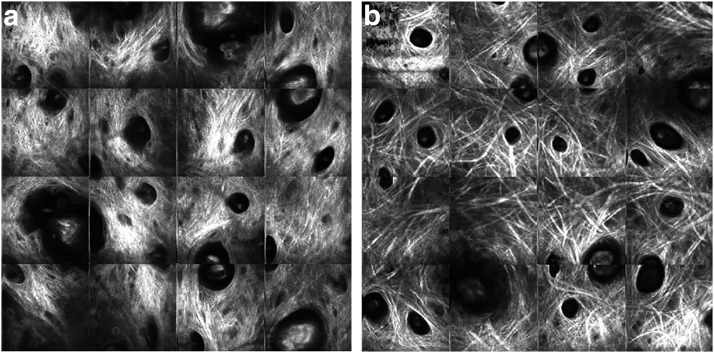
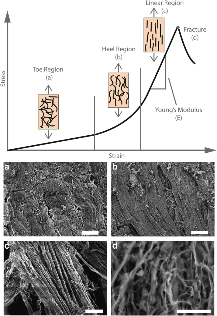
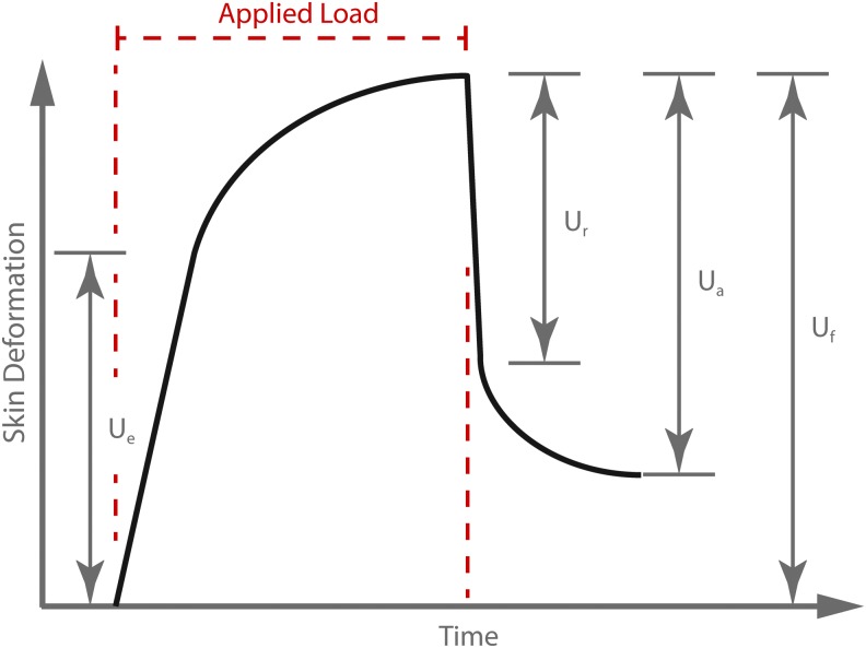
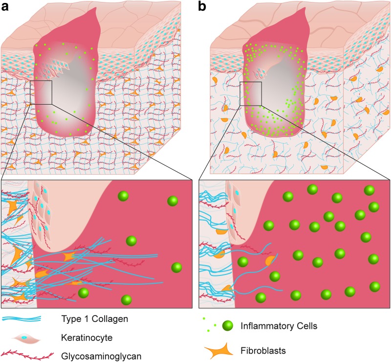
Significance: Chronic wounds, such as diabetic foot ulcers, venous stasis ulcers, and pressure ulcers affect millions of Americans each year, and disproportionately afflict our increasingly older population. Older individuals are predisposed to wound infection, repeated trauma, and the development of chronic wounds. However, a complete understanding of how the attributes of aging skin affect the wound healing process has remained elusive. Recent Advances: A variety of studies have demonstrated that the dermal matrix becomes thinner, increasingly crosslinked, and fragmented with advanced age. These structural changes, as well as an increase in cell senescence, result in altered collagen fiber remodeling and increased stiffness. Studies combining mechanical testing with advanced imaging techniques are providing new insights into the relationships between these age-related changes. Emerging research into the mechanobiology of aging and the wound healing process indicate that the altered mechanical environment of aged skin may have a significant effect on age-related delays in healing. Critical Issues: The interpretation and synthesis of clinical studies is confounded by the effects of common comorbidities that also contribute to the development of chronic wounds. A lack of quantitative biomarkers of wound healing and age-related changes makes understanding structure-function relationships during the wound healing process challenging. Future Directions: Additional work is needed to establish quantitative and mechanistic relationships among age-related changes in the skin microstructure, mechanical function, and the cellular responses to wound healing.

Keywords: collagen; mechanics; stiffness.


Conflict of interest statement

No competing financial interests exist. The content of this article was expressly written by the authors listed. No ghostwriters were used to write this article.

Figures

None
Kyle P. Quinn, PhD
Figure 1.
Figure 1.
Schematic of skin sections in the young (a) and the aged (b). The stratum corneum (1) becomes thicker with age. The epidermis (2) thins with age. Older keratinocytes in the stratum basale (3) are less proliferative. Epidermal rete ridges and dermal papillae (4) at the dermal/epidermal junction flatten with age due to retraction of villi. The papillary dermis (5) contains less collagen than the reticular dermis (6), although the collagen density in both layers decreases with age. Collagen (7) in aged skin is more fragmented and clustered than in young skin. Fibroblasts have a lower migratory potential, and, due to the fragmented ECM, develop fewer focal adhesion complexes and experience fewer external forces (8). The different elements in this schematic are not necessarily to scale. ECM, extracellular matrix. Color images are available online.
Figure 2.
Figure 2.
SEM images of the dermis reveal long fibrils of intertwined collagen The network in young skin (a) is relatively uniform and organized, whereas the aged collagen network (b) contains clumped, fragmented fibrils that accumulate with age. Adapted from Fisher et al. with permission.
Figure 3.
Figure 3.
Multiple SHG images (each at 2.4 × 2.4 mm) of the collagen network from the cheek skin of a 20 year old (a) and a 60 year old (b) reveal thicker, coarser collagen bundles in aged skin. Adapted from Yasui et al. SHG, second-harmonic generation.
Figure 4.
Figure 4.
Skin from donors of different ages (a) were analyzed using FTIR to derive an amide I/amide II ratio using nonpolarized (b) and polarized (c) light. The smaller ratio in (c) with increasing age indicates that the orientation of the collagen fibers becomes more parallel to the surface of the skin. Adapted from Nguyen et al. FTIR, fourier transform infrared spectroscopy. Color images are available online.
Figure 5.
Figure 5.
Image of young (a, 4 month) and aged (b, 23 month) mouse skin, obtained with multiphoton microscopy. Red signal is from collagen SHG excited at 855 nm, and collected with a 420 nm shortpass filter. The green signal is TPEF excited at 755 nm, and collected at 525 ± 25 nm. TPEF is detectable from both cellular autofluorescence (non-SHG-producing regions) and from collagen crosslink autofluorescence (SHG-producing regions). Image settings and skin depth were consistent between (a) and (b). Image size 512 × 512 pixels. Scale bar 100 μm. TPEF, two-photon excited fluorescence. Color images are available online.
Figure 6.
Figure 6.
Schematic of how collagen fiber organization relates to the stress–strain curve during tensile loading. The toe region (a) and heel region (b) of the stress–strain curve correspond to a reorientation of the collagen fibrils parallel to the direction of the applied stress (arrows). The collagen fibrils bear the load in the linear region (c), and begin to separate and break (d) as the load surpasses the ultimate tensile strength of the fibrils. The Young's modulus (E) is a measure of the slope of the stress–strain curve in the linear region. SEM images of the collagen network correspond to loading along the indicated regions (a–d) of the stress–strain curve. The scale bars for the SEM images are 20, 20, 20, and 50 μm, respectively. Figure adapted from Yang et al. (2015). SEM, scanning electron microscopy. Color images are available online.
Figure 7.
Figure 7.
Example of the deformation curve of skin produced under torsion or suction testing. Commonly reported parameters from this type of loading include: the immediate extensibility (Ue), the immediate recovery (Ur), the ultimate recovery (Ua), and the ultimate deformation (Uf). Color images are available online.
Figure 8.
Figure 8.
Schematic of a dermal wound in young (a) and aged (b) subjects. Aged skin exhibits decreased GAG content, delayed proliferation and migration, and decreased fibrosis. Inflammatory cells are more abundant in aged granulation tissue in the later stages of healing. GAG, glycosaminoglycan. Color images are available online.
Figure 9.
Figure 9.
Graphical representation of the biological changes in aged skin that cause downstream effects on ECM structure leading to altered mechanical properties. Color images are available online.

References

    1. Tracy LE, Minasian RA, Caterson EJ. Extracellular matrix and dermal fibroblast function in the healing wound. Adv Wound Care (New Rochelle) 2016;5:119–136 - PMC - PubMed
    1. Harn HI, Ogawa R, Hsu CK, Hughes MW, Tang MJ, Chuong CM. The tension biology of wound healing. Exp Dermatol 2019;28:464–471 - PubMed
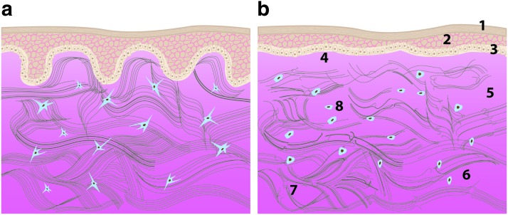
    1. Lavker RM, Zheng PS, Dong G. Morphology of Aged Skin. Dermatol Clin 1986;4:379–389 - PubMed
    1. Grahame R, Holt PJ. The influence of ageing on the in vivo elasticity of human skin. Gerontologia 1969;15:121–139 - PubMed
    1. Rittie L, Fisher GJ. Natural and sun-induced aging of human skin. Cold Spring Harb Perspect Med 2015;5:a015370. - PMC - PubMed

Publication types