

Microsporidial stromal keratitis in a cat
- PMID: 31993317
- PMCID: PMC6976900
- DOI: 10.1016/j.mmcr.2020.01.004
Microsporidial stromal keratitis in a cat
Abstract
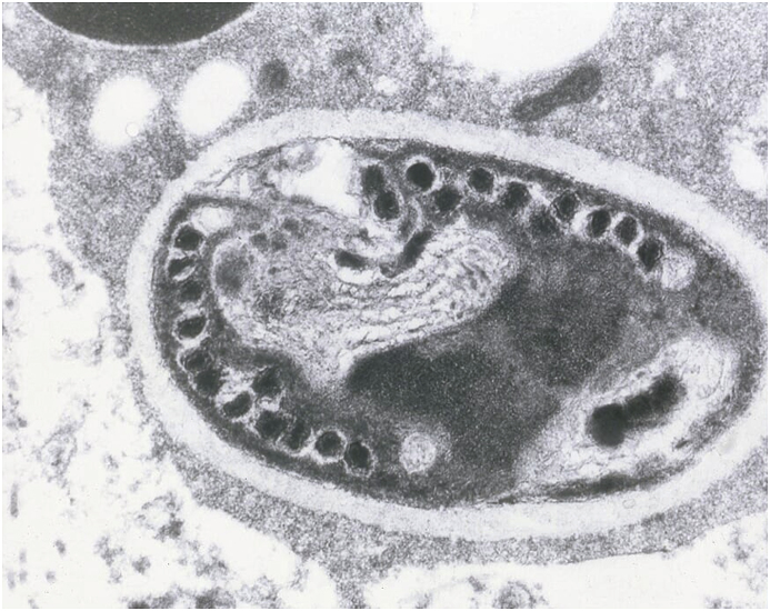
A 12 year-old female spayed felid presented after a 35 day history of right eye pain. On examination, a sub-epithelial opacity was identified in the cornea. A lamellar keratectomy was performed and histopathological analysis revealed low numbers of 2x4um, Gram, Hamatoxylin-eosin and Gomori methanamine-silver positive spores. Transmission electron microscopy found ultrastructural findings consistent with the phylum Microspora. To the author's knowledge, this is only the second case of microsporidial stromal keratitis reported in a felid.
Keywords: Feline; Fungal; Keratitis; Microsporidial; Veterinary.
© 2020 The Authors.
Conflict of interest statement
The authors have no personal or financial conflicts of interest.
Figures


References
-
- Franzen C. Microsporidia: a review of 150 years of research. Open Parasitol. J. 2008;2:1–34.
-
- Didier E., Snowden K., Shadduck J. The biology of microsporidian species infecting mammals. Adv. Parasitol. 1998:279–316. - PubMed
-
- Sabhapandit S., Murthy S., Garg P. Microsporidial stromal keratitis: clinical features, unique diagnostic criteria, and treatment outcomes in a large case series. Cornea. 2016;35(12):1569–1574. - PubMed
-
- Buyukmichi N., Bellhorn R.W., Hunziker J. Encephalitozoon (Nosema) infection of the cornea in a cat. J. Am. Vet. Med. Assoc. 1977;171:355. - PubMed
Publication types
LinkOut - more resources
Full Text Sources
Miscellaneous
