Case Reports
. 2019 Nov 22;3(4):510-516.
doi: 10.1016/j.mayocpiqo.2019.09.005. eCollection 2019 Dec.

Heart Block Caused by Cardiac Metastasis From Merkel Cell Carcinoma: A Case Report

Affiliations
Case Reports

Heart Block Caused by Cardiac Metastasis From Merkel Cell Carcinoma: A Case Report

Nazanin Yeganeh Kazemi et al. Mayo Clin Proc Innov Qual Outcomes. .

Abstract

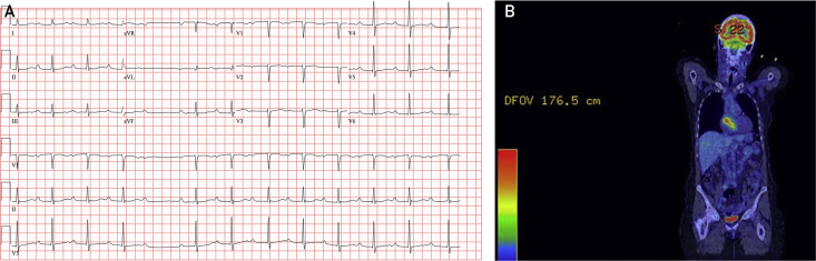
Merkel cell carcinoma (MCC) is a rare, rapidly proliferating skin cancer that commonly metastasizes to regional lymph nodes. We present the case of a 73-year-old woman with a history of MCC and non-Hodgkin B-cell lymphoma who presented with second-degree heart block (Mobitz type II) caused by an interatrial mass. Temporary pacing was required before biopsy, which revealed metastatic MCC. Treatment included permanent pacing, anti-programmed cell death ligand 1 immunotherapy, and radiation to the heart resulting in notable decrease in tumor size and normalized cardiac rhythm.

Keywords: CM, cardiac metastases; CT, computed tomography; FDG, [18F]-fluorodeoxyglucose; MCC, Merkel cell carcinoma; MCPyV, Merkel cell polyomavirus; MRI, magnetic resonance imaging; PD-L1, programmed cell death ligand 1; PET, positron emission tomography.


Figures

Figure 1
Figure 1
A, Electrocardiogram at presentation shows sinus rhythm with prolonged PR interval, narrow QRS complex, and one dropped ventricular beat suggestive of second-degree atrioventricular block, Mobitz type II, which prompted temporary pacing. B, Coronal PET image. The atrial mass exhibits [18F]-fluorodeoxyglucose avidity up to a maximum standardized uptake value of 7.2. Image also demonstrates physiologic uptake in the brain and bladder. PET = positron emission tomography.
Figure 2
Figure 2
Imaging of intracardiac mass and guidance for biopsy. A, Atrial mass on magnetic resonance imaging, 4-chamber view showing the mass circumscribing the aortic root. In this view, the mass measures 35.7 mm. B, Initial transthoracic echocardiogram, Mayo format apical 4-chamber view of mass along interatrial septum. C-E, Imaging for biopsy procedural guidance. C, Anteroposterior projection of flouroscopy. D, Intracardiac echocardiogram showing proximity of the mass to the aortic root. E, Transthoracic echocardiogram showing bioptome at the mass at a level where the left atrium is distal to the mass rather than the aortic root. AV = aortic valve; B = bioptome; ECG = external electrocardiogram wire; ICE = intracardiac echocardiogram probe; LA = left atrium; LV = left ventricle; M = mass; PM = right ventricular pacemaker lead; RA = right atrium; RV = right ventricle; TTE = transthoracic echocardiogram probe.
Figure 3
Figure 3
A-D, Metastatic Merkel cell carcinoma histopathology. Lesional cells demonstrated sheetlike growth with fine nuclear chromatin, scant cytoplasm, and abundant mitotic activity (A and B, hematoxylin-eosin, original magnification ×40 and ×100, respectively). The diagnosis was confirmed with immunoreactivity for CD56 (C, original magnification ×400) and classic perinuclear dotlike reactivity with CK20 (D, original magnification ×400). E, Scan obtained at time of admission when the patient presented with heart block, demonstrating the size and location of the mass around the aortic root. F, Scan 2 months after starting treatment.

References

    1. Paulson K.G., Park S.Y., Vandeven N.A., et al. Merkel cell carcinoma: current US incidence and projected increases based on changing demographics. J Am Acad Dermatol. 2018;78(3):457–463.e2. - PMC - PubMed
    1. Heath M., Jaimes N., Lemos B., et al. Clinical characteristics of Merkel cell carcinoma at diagnosis in 195 patients: the AEIOU features. J Am Acad Dermatol. 2008;58(3):375–381. - PMC - PubMed
    1. Albores-Saavedra J., Batich K., Chable-Montero F., Sagy N., Schwartz A.M., Henson D.E. Merkel cell carcinoma demographics, morphology, and survival based on 3870 cases: a population based study. J Cutan Pathol. 2010;37(1):20–27. - PubMed
    1. Wong H.H., Wang J. Merkel cell carcinoma. Arch Pathol Lab Med. 2010;134(11):1711–1716. - PubMed
    1. Warner T.F., Uno H., Hafez G.R., et al. Merkel cells and Merkel cell tumors: ultrastructure, immunocytochemistry and review of the literature. Cancer. 1983;52(2):238–245. - PubMed

Publication types