. 2020 May 8;29(7):1121-1131.
doi: 10.1093/hmg/ddaa018.

A mouse model for kinesin family member 11 (Kif11)-associated familial exudative vitreoretinopathy

Affiliations

A mouse model for kinesin family member 11 (Kif11)-associated familial exudative vitreoretinopathy

Yanshu Wang et al. Hum Mol Genet. .

Abstract

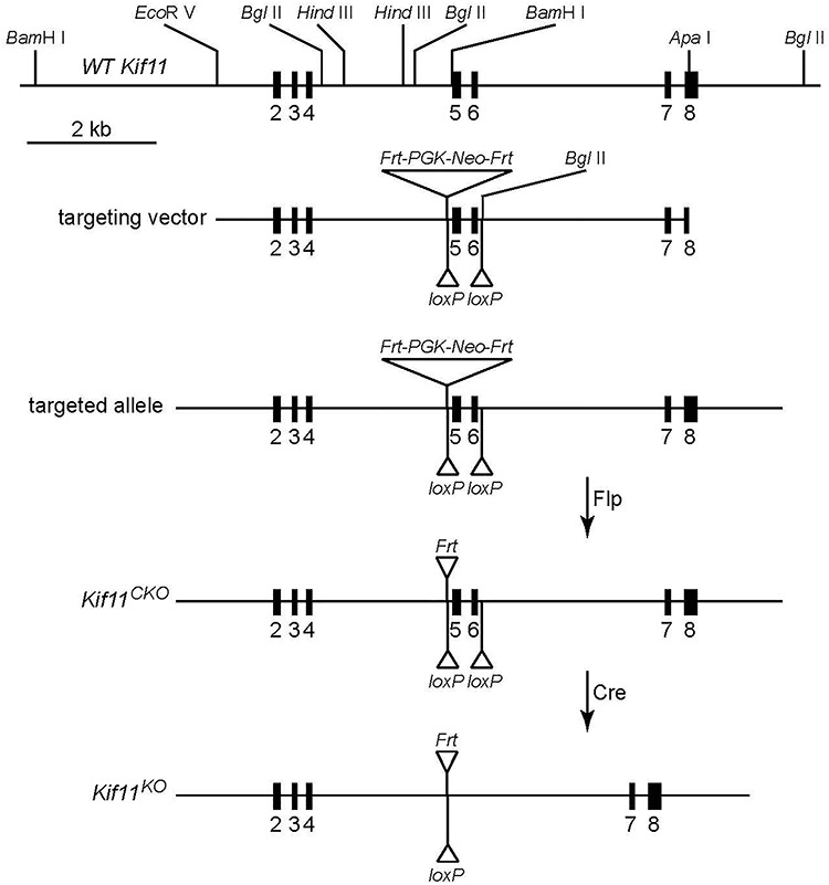
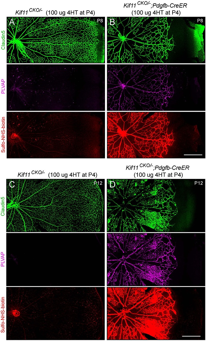
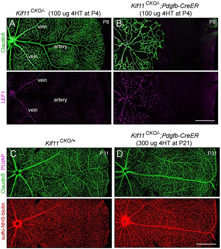
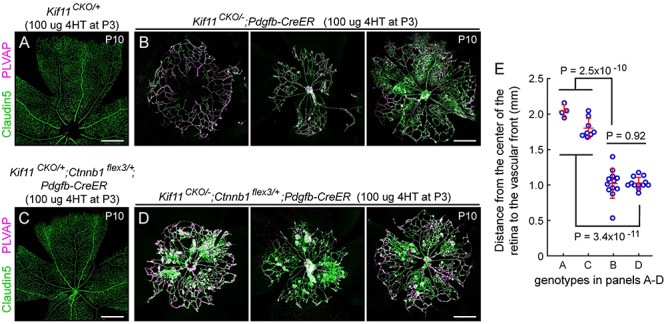
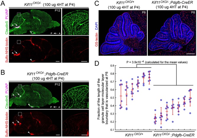
During mitosis, Kif11, a kinesin motor protein, promotes bipolar spindle formation and chromosome movement, and during interphase, Kif11 mediates diverse trafficking processes in the cytoplasm. In humans, inactivating mutations in KIF11 are associated with (1) retinal hypovascularization with or without microcephaly and (2) multi-organ syndromes characterized by variable combinations of lymphedema, chorioretinal dysplasia, microcephaly and/or mental retardation. To explore the pathogenic basis of KIF11-associated retinal vascular disease, we generated a Kif11 conditional knockout (CKO) mouse and investigated the consequences of early postnatal inactivation of Kif11 in vascular endothelial cells (ECs). The principal finding is that postnatal EC-specific loss of Kif11 leads to severely stunted growth of the retinal vasculature, mildly stunted growth of the cerebellar vasculature and little or no effect on the vasculature elsewhere in the central nervous system (CNS). Thus, in mice, Kif11 function in early postnatal CNS ECs is most significant in the two CNS regions-the retina and cerebellum-that exhibit the most rapid rate of postnatal growth, which may sensitize ECs to impaired mitotic spindle function. Several lines of evidence indicate that these phenotypes are not caused by reduced beta-catenin signaling in ECs, despite the close resemblance of the Kif11 CKO phenotype to that caused by EC-specific reductions in beta-catenin signaling. Based on prior work, defective beta-catenin signaling had been the only known mechanism responsible for monogenic human disorders of retinal hypovascularization. The present study implies that retinal hypovascularization can arise from a second and mechanistically distinct cause.


Figures

Figure 1
Figure 1
Structure of the Kif11 CKO allele. Shown from top to bottom are the region of the WT Kif11 allele encompassing exons 2–8, the ES cell targeting vector, the targeted allele, the CKO allele and the KO allele following the Cre-mediated recombination of the CKO allele.
Figure 2
Figure 2
Severely retarded retinal vascular growth following the early postnatal EC-specific knockout of Kif11. Retinal flatmounts were prepared at P8 or P12 from littermate control Kif11CKO/− mice (A,C) and Kif11CKO/−;Pdgfb-CreER mice (B,D) following the treatment with 100 μg 4HT at P4. Each image shows one retinal quadrant, with the optic disc at the left and the retinal periphery at the right. Loss of Kif11 leads to retarded vascular development and reduced vascular coverage of the retinal surface. At both ages, Kif11CKO/−;Pdgfb-CreER retinas show increased expression of PLVAP and increased Sulfo-NHS-biotin leakage. Scale bars, 500 μm.
Figure 3
Figure 3
EC-specific knockout of Kif11 leads to continued EC accumulation of LEF1 during development and no effect of post-mitotic knockout on vascular permeability. (A,B) Retinal flatmounts were prepared at P8 from littermate control Kif11CKO/− mice (A) and Kif11CKO/−;Pdgfb-CreER mice (B) following the treatment with 100 μg 4HT at P4. Each image shows one retinal quadrant, with the optic disc at the left and the retinal periphery at the right. LEF1, a marker of beta-catenin signaling, accumulates in the nuclei of vein and capillary ECs but not in arterial ECs in control retinas and in all ECs in mutant retinas. (C,D) Retinal flatmounts were prepared at P31 from littermate control Kif11CKO/+ mice (C) and Kif11CKO/−;Pdgfb-CreER mice (D) following the treatment with 300 μg 4HT at P21. Each image shows one retinal quadrant as in (A) and (B). Loss of Kif11 has no effect on vascular architecture, expression of Claudin5, suppression of PLVAP or BRB integrity, as seen by the absence of Sulfo-NHS-biotin leakage from the intravascular space into the retinal parenchyma. See Supplementary Material Figure S1 for an analysis of Cre-mediated recombination at P21. Scale bars, 500 μm.
Figure 4
Figure 4
Postnatal EC-specific stabilization of beta-catenin does not rescue the Kif11 retinal vascular phenotype. (A,B) Retinal flatmounts were prepared at P10 from control Kif11CKO/+ mice (A) and Kif11CKO/−;Pdgfb-CreER mice (B; three examples shown) following the treatment with 100 μg 4HT at P3 to assess the Kif11 retinal vascular phenotype. (C,D) From the same set of crosses, retinal flatmounts were prepared at P10 from control Kif11CKO/+;Ctnnb1flex3/+;Pdgfb-CreER mice (C) and Kif11CKO/−;Ctnnb1flex3/+;Pdgfb-CreER mice (D; three examples shown) following the treatment with 100 μg 4HT at P3. Retinal vascular development in both sets of control mice is indistinguishable from WT. Beta-catenin stabilization following the Cre-mediated recombination of Ctnnb1flex3/+ fails to rescue the Kif11 retinal vascular phenotype. In (A) and (C), each image shows one retinal quadrant, with the optic disc at the bottom and the retinal periphery at the top; in (B) and (D), each image is centered on the optic disc. All images in (A–D) are at the same magnification. Scale bars, 500 μm. (E) Quantification of the radial distance from the optic disc to the vascular front at P10 for individual retinal quadrants for the genotypes shown in panels A–D. Plots show mean ± standard deviation.
Figure 5
Figure 5
Retarded density of cerebellar vasculature following the early postnatal EC-specific knockout of Kif11. (A,B) Sagittal brain sections were prepared at P12 from control Kif11CKO/− mice (A) and Kif11CKO/−;Pdgfb-CreER mice (B) following the treatment with 100 μg 4HT at P4. The region of the dorsal cerebellum enclosed by the white square is enlarged at right. Scale bars, 1 mm (left images) and 200 μm (right images). Except for modestly reduced vascular density in the cerebellum (inset at right), brain size and vascular density are unaffected by EC-specific loss of Kif11. Sulfo-NHS-biotin leakage is seen in the choroid plexus (arrow) and in the circumventricular organs (area postrema and median eminence; arrowheads) as expected but is not observed elsewhere in the brain. (C) Sagittal brain sections were prepared at P8 from control Kif11CKO/+ mice and Kif11CKO/−;Pdgfb-CreER mice, following the treatment with 100 μg 4HT at P4. Scale bar, 500 μm. (D) With early postnatal loss of Kif11, vascular density in the P8 cerebellum is reduced, as determined by quantifying the fraction of the molecular layer/granule cell layer border that is occupied by blood vessels in six control Kif11CKO/+ and seven Kif11CKO/−;Pdgfb-CreER P8 mice. For each mouse, seven folia were quantified from a single confocal stack of the cerebellum [examples in (C)]. The plot in (D) shows mean ± standard deviation for each mouse.

References

    1. Gilmour D.F. (2015) Familial exudative vitreoretinopathy and related retinopathies. Eye (Lond.), 29, 1–14. - PMC - PubMed
    1. Sun Y. and Smith L.E.H. (2018) Retinal vasculature in development and diseases. Annu. Rev. Vis. Sci., 4, 101–122. - PMC - PubMed
    1. Nikopoulos K., Venselaar H., Collin R.W., Riveiro-Alvarez R., Boonstra F.N., Hooymans J.M., Mukhopadhyay A., Shears D., van Bers M., de Wijs I.J. et al. (2010) Overview of the mutation spectrum in familial exudative vitreoretinopathy and Norrie disease with identification of 21 novel variants in FZD4, LRP5, and NDP. Hum. Mutat., 31, 656–666. - PubMed
    1. Rao F.Q., Cai X.B., Cheng F.F., Cheng W., Fang X.L., Li N., Huang X.F., Li L.H. and Jin Z.B. (2017) Mutations in LRP5,FZD4, TSPAN12, NDP, ZNF408, or KIF11 genes account for 38.7% of Chinese patients with familial exudative vitreoretinopathy. Invest. Ophthalmol. Vis. Sci., 58, 2623–2629. - PubMed
    1. Tang M., Sun L., Hu A., Yuan M., Yang Y., Peng X. and Ding X. (2017) Mutation spectrum of the LRP5, NDP, and TSPAN12 genes in Chinese patients with familial exudative vitreoretinopathy. Invest. Ophthalmol. Vis. Sci., 58, 5949–5957. - PubMed

Publication types