

A morphologic and molecular reappraisal of myoepithelial tumors of soft tissue, bone, and viscera with EWSR1 and FUS gene rearrangements
- PMID: 31994243
- PMCID: PMC7170037
- DOI: 10.1002/gcc.22835
A morphologic and molecular reappraisal of myoepithelial tumors of soft tissue, bone, and viscera with EWSR1 and FUS gene rearrangements
Abstract
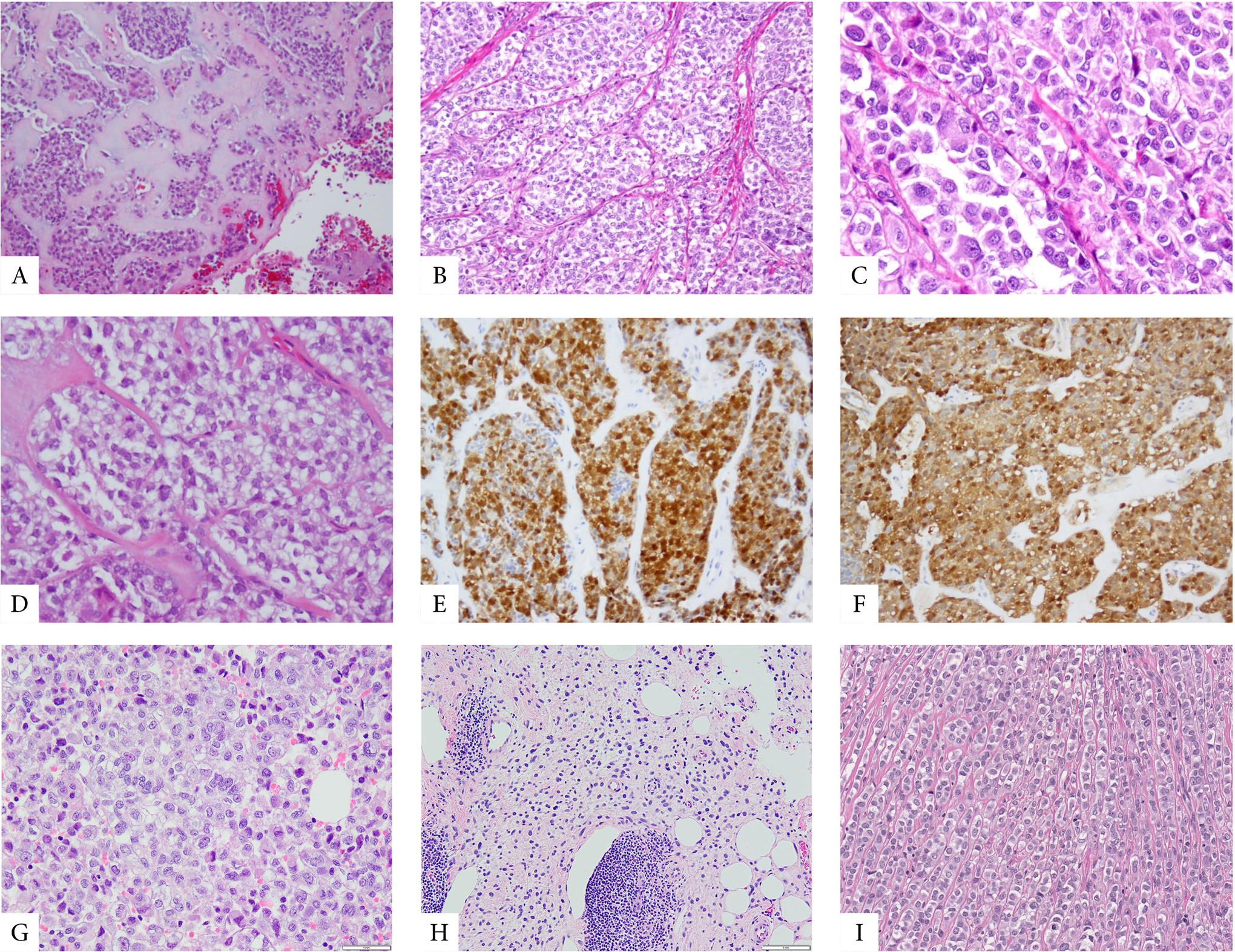
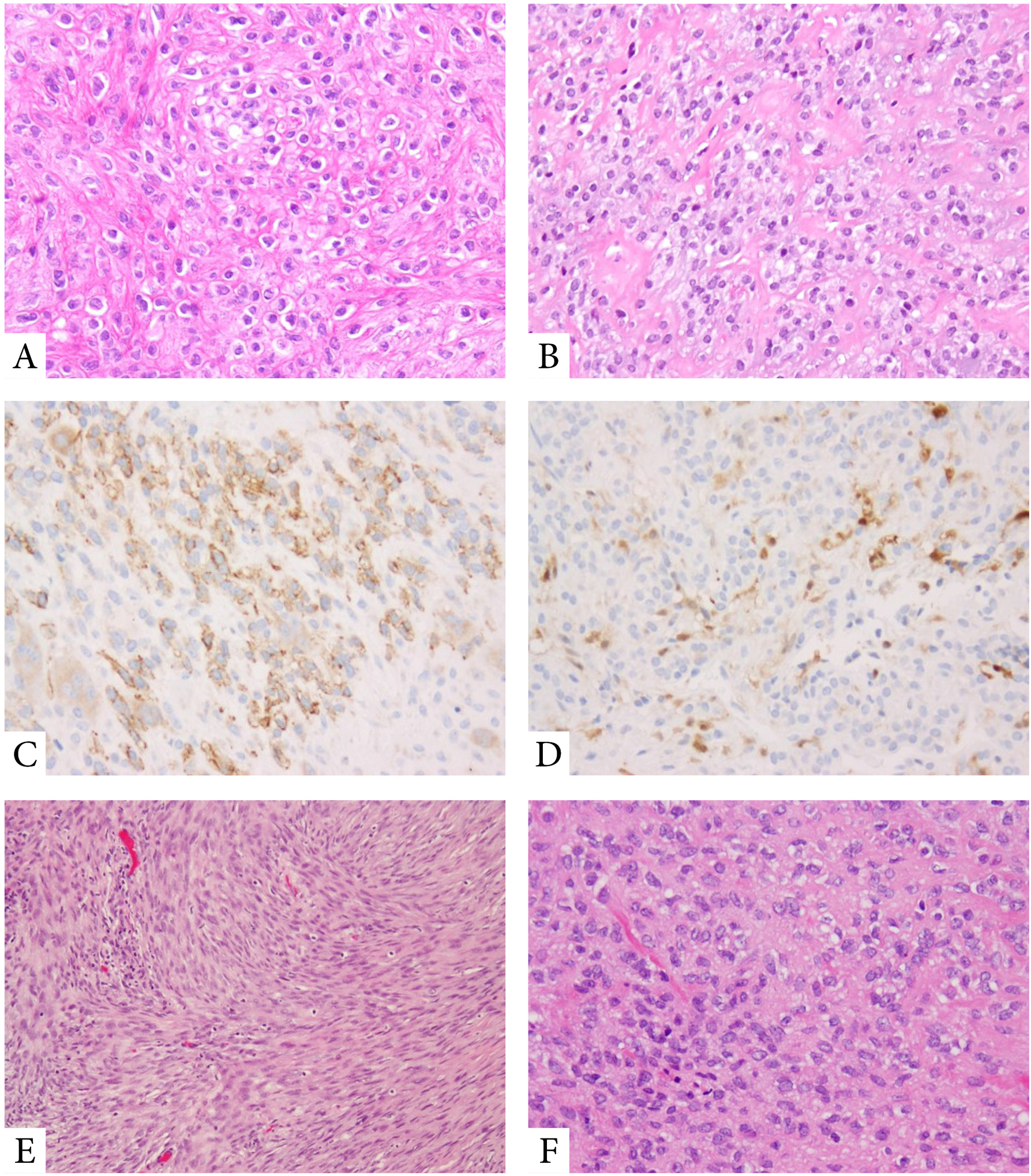
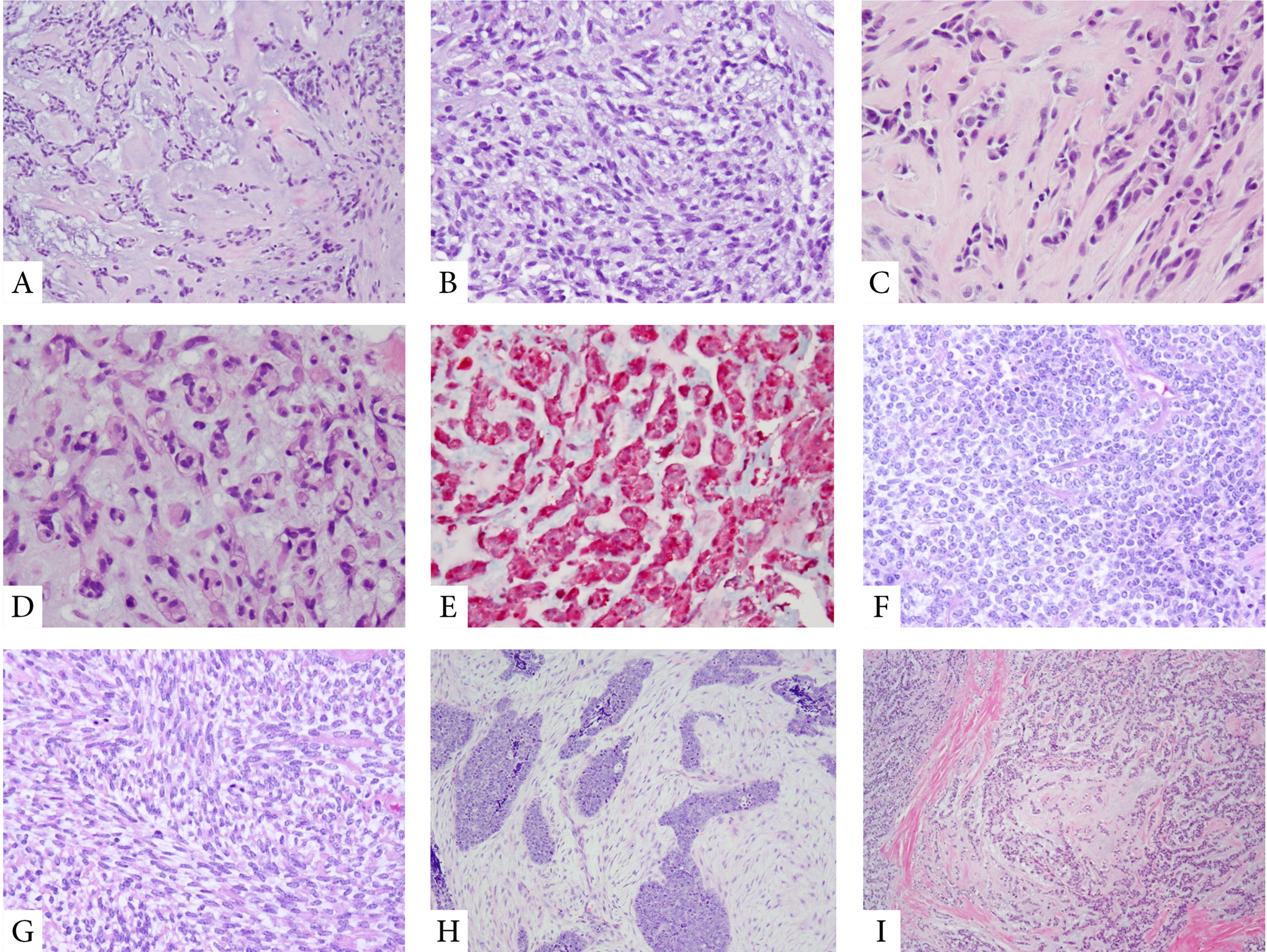
Myoepithelial tumors (MET) represent a clinicopathologically heterogeneous group of tumors, ranging from benign to highly aggressive lesions. Although MET arising in soft tissue, bone, or viscera share morphologic and immunophenotypic overlap with their salivary gland and cutaneous counterparts, there is still controversy regarding their genetic relationship. Half of MET of soft tissue and bone harbor EWSR1 or FUS related fusions, while MET arising in the salivary gland and skin often show PLAG1 and HMGA2 gene rearrangements. Regardless of the site of origin, the gold standard in diagnosing a MET relies on demonstrating its "myoepithelial immunophenotype" of positivity for EMA/CK and S100 protein or GFAP. However, the morphologic spectrum of MET in soft tissue and bone is quite broad and the above immunoprofile is nonspecific, being shared by other pathogenetically unrelated neoplasms. Moreover, rare MET lack a diagnostic immunoprofile but shows instead the characteristic gene fusions. In this study, we analyzed a large cohort of 66 MET with EWSR1 and FUS gene rearrangements spanning various clinical presentations, to better define their morphologic spectrum and establish relevant pathologic-molecular correlations. Genetic analysis was carried out by FISH for EWSR1/FUS rearrangements and potential partners, and/or by targeted RNA sequencing. Then, 82% showed EWSR1 rearrangement, while 18% had FUS abnormalities. EWSR1-POU5F1 occurred with predilection in malignant MET in children and young adults and these tumors had nested epithelioid morphology and clear cytoplasm. In contrast, EWSR1/FUS-PBX1/3 fusions were associated with benign and sclerotic spindle cell morphology. Tumors with EWSR1-KLF17 showed chordoma-like morphology. Our results demonstrate striking morphologic-molecular correlations in MET of bone, soft tissue and viscera, which might have implications in their clinical behavior.
Keywords: EWSR1; FUS; PBX1; PBX3; POU5F1; myoepithelial tumors.
© 2020 Wiley Periodicals, Inc.
Conflict of interest statement
Figures

References
-
- Hornick JL, Fletcher CD. Myoepithelial tumors of soft tissue: a clinicopathologic and immunohistochemical study of 101 cases with evaluation of prognostic parameters. Am J Surg Pathol. 2003;27:1183–1196. - PubMed
-
- Song W, Flucke U, Suurmeijer AJH. Myoepithelial Tumors of Bone. Surg Pathol Clin. 2017;10:657–674. - PubMed
-
- Katabi N, Gomez D, Klimstra DS, et al. Prognostic factors of recurrence in salivary carcinoma ex pleomorphic adenoma, with emphasis on the carcinoma histologic subtype: a clinicopathologic study of 43 cases. Hum Pathol. 2010;41:927–934. - PubMed
Publication types
MeSH terms
Substances
Grants and funding
LinkOut - more resources
Full Text Sources
Medical
Research Materials
Miscellaneous
