Comparative Study
. 2020 Jan 29;14(1):e0008039.
doi: 10.1371/journal.pntd.0008039. eCollection 2020 Jan.

Comparative specificity and sensitivity of NS1-based serological assays for the detection of flavivirus immune response

Affiliations
Comparative Study

Comparative specificity and sensitivity of NS1-based serological assays for the detection of flavivirus immune response

Erick Mora-Cárdenas et al. PLoS Negl Trop Dis. .

Abstract

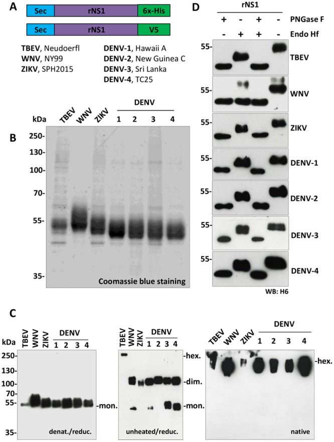
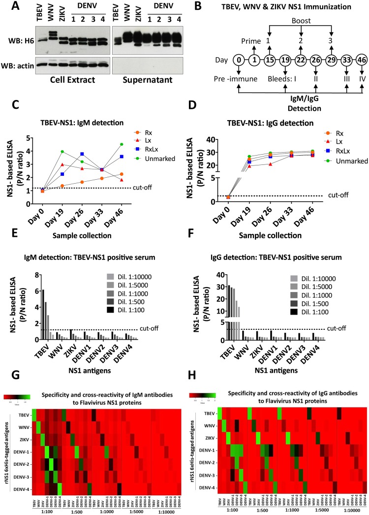
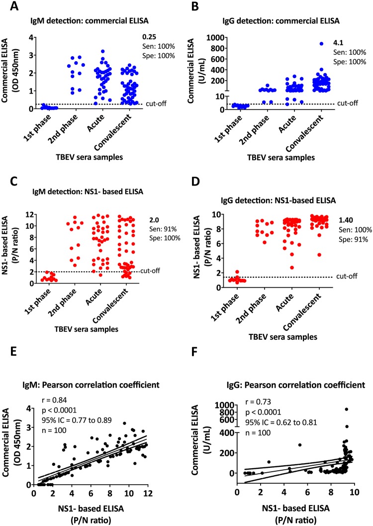
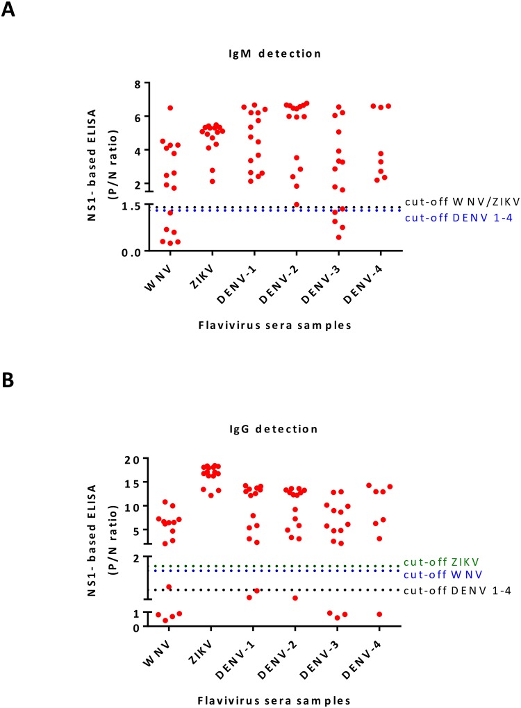
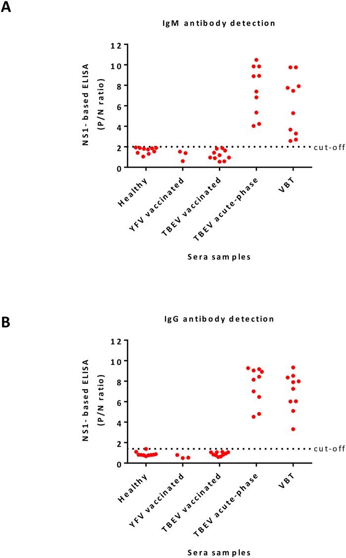
Flaviviruses are relevant animal and human pathogens of increasing importance worldwide. The similarities of the initial clinical symptoms and the serological cross-reactivity of viral structural antigens make a laboratory diagnosis of flavivirus infection problematic. The main aim of the present study was the comparative specificity and sensitivity analysis of the non-structural protein NS1 as an antigen to detect flavivirus antibodies in sera from exposed individuals. A strategy for the purification of native recombinant non-structural protein 1 of representative flaviviruses including tick-borne encephalitis, West Nile, Zika and dengue virus was developed. The immunological properties of the purified antigens were analyzed using sera of immunized mice and of infected individuals in comparison with standard commercial assays. Recombinant NS1 protein was confirmed as a valuable option for the detection of flavivirus antibodies with reduced cross-reactivity and high sensitivity offering additional advantages for the detection of vaccine breakthrough cases.


Conflict of interest statement

The authors have declared that no competing interests exist.

Figures

Fig 1
Fig 1. Cloning, expression, and purification of rNS1.
A) Schematic representation of expression constructs pcDNA3.1-sec-NS1-6xHis (upper) and pcDNA3.1-sec-NS1-V5 (lower). Sec corresponds to an immunoglobulin leader sequence at the N-term; rNS1, recombinant nonstructural protein 1; 6xHis and V5 are tags cloned at the C-term for protein purification and mice immunization, respectively. Reference strains are also indicated. B) Coomassie blue staining of purified proteins: 2 μg of purified NS1 proteins were loaded for each virus. C) Western blot analysis of purified rNS1. Left panel, WB of purified rNS1 proteins diluted in reducing Laemmli sample buffer (LB) and denatured by boiling using an anti-6xHis-tag mAb (WB: H6). Middle panel, WB of rNS1 performed in non-denaturing/non-reducing conditions (without heating the proteins and LB without 2-βME). Right panel, WB of purified rNS1 proteins analyzed by western blot under native conditions (without heating the proteins; without 2-βME and without SDS in the LB buffer, gel, and running buffer). rNS1 monomers (mon), dimers (dim), and hexamers (hex) are indicated. D) Endoglycosidase analysis of purified rNS1. All rNS1 proteins were treated (+)/or not (-) with PNGase F and/or Endo Hf enzymes for 1.5 h at 37 °C. WB analysis was assessed by 10% SDS-PAGE under standard conditions using an anti-6xHis-tag mAb (WB: H6).
Fig 2
Fig 2. Validation of rNS1 antigens by ELISA in immunized mice.
A) Expression and secretion of V5-tagged recombinant NS1 proteins of TBEV, WNV, ZIKV, and DENV1-4 in cell extract and supernatant of transfected HEK293T cells. Actin blots were included as a loading control for cell extracts and of clean supernatants. SDS PAGE was conducted in denaturing/reducing conditions. B) Schematic representation of the protocol followed for the immunization of BALB/c mice by gene gun technology. Numbers indicate the day when the prime-boost immunization was performed and the day of collection. C and D) TBEV rNS1 IgM/IgG ELISA with sera from four different mice immunized with TBEV-NS1 (mice were differentiated with a mark in the right ear (Rx), left (Lx), both ears (RxLx) or without the mark (unmarked)). Detection of IgM/IgG antibodies was performed at day 0, 19, 26, 33 and 46. E and F) TBEV, WNV, ZIKV, and DENV1-4 IgM/IgG rNS1 ELISA with sera from TBEV-NS1 immunized mice. Sera from day 46 post-immunization were diluted in blocking solution (2% milk in PBS) as indicated. G and H) Heatmaps of the IgM/IgG cross-reactivity. Several dilutions of each serum sample were analyzed in plates coated with homologous and heterologous rNS1 6xHis-tagged antigens. * each ELISA result includes the average of two biological replicates.
Fig 3
Fig 3. Detection of IgM/IgG antibodies from TBEV infected individuals.
A and B) Detection of IgM (A) IgG (B) antibodies by commercial ELISA. IgM results are reported as OD450, while IgG results are reported as U/mL. Cut-off values for IgM (0.25) and IgG (4.1) were calculated according to the manufacturer’s instructions. C and D) Detection of IgM (C) IgG (D) antibodies by TBEV rNS1-based ELISA. Optimal cut-off values of the P/N ratio (OD450 of test specimen divided by the mean OD450 of negative control specimens) were calculated based on the comparative receiver operating characteristic (ROC) curve analysis. Cut-off values for IgM and IgG fell at 2.0 and 1.40, respectively. E and F) Correlation between commercial and TBEV rNS1-based ELISA assays. The two-tailed Pearson’s correlation value (r) was calculated for IgM and IgG values. A P value of <0.0001 rejected the null hypothesis that there exists no correlation between commercial and rNS1-based ELISA methods. 95% Interval Confidence (IC) value is also indicated and showed in dotted lines above and under the linear correlation. 100 sera samples (n) from different phases of TBEV infection were included in the analysis. * each ELISA result includes the average of two biological replicates.
Fig 4
Fig 4. Detection of IgM/IgG antibodies from WNV, ZIKV and DENV 1–4 infected individuals.
A and B) Detection of IgM (A) IgG (B) antibodies by rNS1-based ELISA. Each group of RT-PCR confirmed patients were screened for the presence of IgM/IgG antibodies by rNS1-based ELISA tests using specific purified rNS1 antigens. Optimal cut-off values of P/N ratios were calculated based on ROC curve analysis. Each cut-off was selected based on the P/N ratio value, which gave 100% sensitivity and specificity. * each ELISA result includes the average of two biological replicates.
Fig 5
Fig 5. Differential serological diagnosis of TBEV.
A and B) Detection of IgM (A) IgG (B) antibodies by TBEV rNS1-based ELISA using sera samples from five different groups of sera samples: 10 sera samples of healthy individuals from non-endemic TBEV regions and 3 sera from YFV vaccinated individuals (TBEV non-endemic areas) were used as controls. TBEV vaccinated group corresponds to 10 sera from individuals who got the vaccine (a suspension of purified TBE inactivated virus). The acute group corresponds to individuals with acute TBEV infection. The vaccine breakthrough (VBT) group corresponds to individuals who got the vaccine and were also infected with TBEV. * each ELISA result includes the average of two biological replicates.

References

    1. Leta S, Beyene TJ, De Clercq EM, Amenu K, Kraemer MUG, Revie CW. Global risk mapping for major diseases transmitted by Aedes aegypti and Aedes albopictus. Int J Infect Dis. 2018;67:25–35. 10.1016/j.ijid.2017.11.026 - DOI - PMC - PubMed
    1. Pandit PS, Doyle MM, Smart KM, Young CCW, Drape GW, Johnson CK. Predicting wildlife reservoirs and global vulnerability to zoonotic Flaviviruses. Nat Commun. 2018;9(1):5425 10.1038/s41467-018-07896-2 - DOI - PMC - PubMed
    1. Williams DT, Mackenzie JS, Bingham J. Flaviviruses In: Zimmerman JJ, Karriker LA, Ramirez A, Schwartz KJ, Stevenson GW, Zhang J, editors. Diseases of swine: John Wiley & Sons, Inc; 2019.
    1. Carletti T, Zakaria MK, Marcello A. The host cell response to tick-borne encephalitis virus. Biochem Biophys Res Commun. 2017;492(4):533–40. Epub 2017/02/09. 10.1016/j.bbrc.2017.02.006 . - DOI - PubMed
    1. Salimi H, Cain MD, Klein RS. Encephalitic Arboviruses: Emergence, Clinical Presentation, and Neuropathogenesis. Neurotherapeutics. 2016;13(3):514–34. 10.1007/s13311-016-0443-5 - DOI - PMC - PubMed

Publication types