. 2020 Mar 3;28(3):281-289.e3.
doi: 10.1016/j.str.2020.01.003. Epub 2020 Jan 28.

MyD88 Death-Domain Oligomerization Determines Myddosome Architecture: Implications for Toll-like Receptor Signaling

Affiliations

MyD88 Death-Domain Oligomerization Determines Myddosome Architecture: Implications for Toll-like Receptor Signaling

Martin C Moncrieffe et al. Structure. .

Abstract

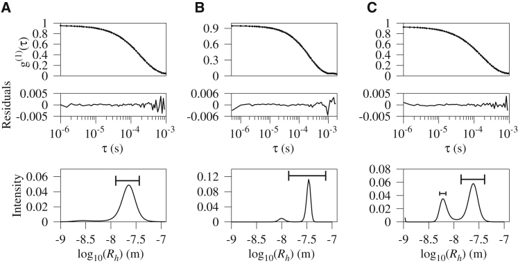
Toll-like receptors (TLRs) are pivotal in triggering the innate immune response to pathogen infection. Ligand binding induces receptor dimerization which facilitates the recruitment of other post-receptor signal transducers into a complex signalosome, the Myddosome. Central to this process is Myeloid differentiation primary response 88 (MyD88), which is required by almost all TLRs, and signaling is thought to proceed via the stepwise, sequential assembly of individual components. Here, we show that the death domains of human MyD88 spontaneously and reversibly associate to form helical filaments in vitro. A 3.1-Å cryoelectron microscopy structure reveals that the architecture of the filament is identical to that of the 6:4 MyD88-IRAK4-IRAK2 hetero-oligomeric Myddosome. Additionally, the death domain of IRAK4 interacts with the filaments to reconstitute the non-stoichiometric 6:4 MyD88-IRAK4 complex. Together, these data suggest that intracellularly, the MyD88 scaffold may be pre-formed and poised for recruitment of IRAKs on receptor activation and TIR engagement.

Keywords: IRAK; MyD88; Myddosome; TIRFM; TLR signaling; cryo-EM; light-sheet microscopy.


Conflict of interest statement

Declaration of Interests The authors declare no competing interests.

Figures

Figure 1
Figure 1
Sedimentation Velocity and Equilibrium Measurement of the Homo-oligomeric MyD88 Death-Domain Complex (A) Velocity data, residuals after fitting, and the c(S) distribution for the hMyD88 death-domain complex. The c(S) distribution is broad and has a peak at 30 S. (B) Sedimentation equilibrium data recorded at 250 nm and 280 nm and rotor speeds of 3,000 and 4,000 rpm. The recovered molecular weight of the hMyD88DD oligomer from a global analysis of these data was 5.2 MDa.
Figure 2
Figure 2
Cryo-EM Structure of the hMyD88DD Filament at 3.1 Å Resolution (A) Typical cryo-EM image of the hMyD88DD filaments, which have lengths ranging from approximately 10 to 1,000 nm. (B–F) Two views of the reconstructed hMyD88DD complex (B and C) with the latter looking down the helical axis and showing the approximately 16-Å cavity that spans the filament. The long helix (H6) at the C terminus of the hMyD88DD is clearly visible. The quality of the cryo-EM map allowed unambiguous fitting of atomic models of hMyD88DD monomers into the reconstructed map. Typical side-chain densities of some residues in the hydrophobic region surrounding W78 (D) and the sixth α-helix, H6 (E). Cartoon representation of a 13-subunit segment of the hMyD88DD monomers superimposed onto the reconstructed cryo-EM map (F).
Figure 3
Figure 3
Structure of the hMyD88DD Filament (A) A segment composed of 13 hMyD88DD subunits (M1–M13). The 13 death-domain subunits are assembled in layers and each layer is colored differently. (B) View along the helical axis of the hMyD88DD filament and some inter-subunit contacts. (C) Superposition of the heterotrimeric hMyD88DD (light gray), IRAK4DD (medium gray), and IRAK2DD (dark gray) Myddosome onto the hMyD88DD filament. The MyD88 layer of the heterotrimer superimposes onto the helical filament with an RMSD of 0.76 Å. (D) Representation of the different interfaces present in the hMyD88DD helical filament. The interaction between the i+1 and i+4 subunits generates a type I interface while that between the i and i+4 subunits generates a type II interface. The type III interface, which occurs at the intersection of type I and II interfaces, is represented by the interaction between the i+3 and i+4 subunits. Red and black clasps indicate where the M3 and M7 subunits fit when the two-dimensional hexagons are wrapped to represent the helical filament.
Figure 4
Figure 4
IRAK4DD Associates with the hMyD88DD Filament The upper and middle panels show plots of the autocorrelation as a function of time and residuals after fitting while the bottom panel shows a plot of intensity as a function of hydrodynamic radius for the hMyD88DD death-domain filament at 34 μM (A) and 45 μM (B), with the length of each bar representing the average filament length. At the highest concentration, the complex has a hydrodynamic radius of approximately 31 ± 9 nm and the distribution shifts to larger oligomers. The addition of IRAK4DD death domain (C) results in the formation of the previously characterized hMyD88DD-IRAK4DD Myddosome (blue).
Figure 5
Figure 5
Full-Length MyD88 Self-associates to Produce a Pre-Myddosome Scaffold In Vivo (A and B) Representative TIRFM (A) and light-sheet (B) images of GFP-MyD88 expression in unstimulated virally transduced MyD88−/− bone-derived macrophage (n = 31). (C) shows a typical TIRFM image after stimulation with LPS (n = 8). Fitting the intensity distribution of the oligomeric GFP-MyD88 species using a box size of 100 pixels to a Gaussian allows determination of the peak intensity, which is used along with that of monomeric GFP-MyD88 (Figures S6C and S6D) to estimate the number of associating MyD88 monomers. For TIRFM and light-sheet illumination in both unstimulated and LPS-stimulated cells, the estimated number of MyD88 monomers was 1 and 5 ± 1, respectively. The scale bars represent 5 μm.
Figure 6
Figure 6
Model of TLR Signaling in which the TIR Domain of hMyD88 Acts as a Binary Switch (A–D) (A) MyD88 contains an N-terminal death domain (DD) and a C-terminal TIR domain, which are linked by an intermediate domain (ID) that is devoid of ordered secondary structure. Cytosolic MyD88 reversibly self-associates via its DD to form an inactive two-layered (M1–M3 and M4–M6) hexameric scaffold that is incapable of producing a signaling Myddosome because the M3–M6 surface, which is required for IRAK4 DD interaction, is obscured by the TIR domain. This inactive conformation could be caused by the TIR domain of the M3 MyD88 subunit (black) from the first layer blocking the M4 and M6 IRAK4 interaction surfaces (B) or, alternatively, weak association between the M3 and M4 TIR domains, for example as shown in (C). Microbe-associated molecular pattern and coreceptor binding—where required—to the receptor ectodomain enable dimerization and activation of receptor TIR domains, which associate directly via intra-strand contacts (D). This receptor TIR dimer interacts strongly with the hMyD88 TIR domain via inter-strand contacts directly or via MAL/TIRAP, and this relieves the inhibition of the TIR domain and allows recruitment of the IRAK4 and IRAK2 onto the hMyD88 scaffold where signal transduction proceeds by phosphorylation.

Comment in

  • Cobbling Together the Myddosome.
    Snyder MLD, Snyder GA. Snyder MLD, et al. Structure. 2020 Jun 2;28(6):598-600. doi: 10.1016/j.str.2020.05.006. Structure. 2020. PMID: 32492411

References

    1. Adams P.D., Afonine P.V., Bunkóczi G., Chen V.B., Davis I.W., Echols N., Headd J.J., Hung L.W., Kapral G.J., GrosseKunstleve R.W. PHENIX: a comprehensive Python-based system for macromolecular structure solution. Acta Crystallogr. D Biol. Crystallogr. 2010;66:213–221. - PMC - PubMed
    1. Akira S., Uematsu S., Takeuchi O. Pathogen recognition and innate immunity. Cell. 2006;124:783–801. - PubMed
    1. Avbelj M., Wolz O.O., Fekonja O., Benčina M., Repič M., Mavri J., Krüger J., Schärfe C., Delmiro Garcia M., Panter G. Activation of lymphoma-associated MyD88 mutations via allostery-induced TIR-domain oligomerization. Blood. 2014;124:3896–3904. - PMC - PubMed
    1. Beutler B. Inferences, questions and possibilities in Toll-like receptor signalling. Nature. 2004;430:257–263. - PubMed
    1. Bray D., Levin M.D., MortonFirth C.J. Receptor clustering as a cellular mechanism to control sensitivity. Nature. 1998;393:85–88. - PubMed

Publication types

MeSH terms

Substances

LinkOut - more resources