. 2020 Jan 29;20(1):22.
doi: 10.1186/s12866-019-1635-z.

A mouse ear skin model to study the dynamics of innate immune responses against Staphylococcus aureus biofilms

Affiliations

A mouse ear skin model to study the dynamics of innate immune responses against Staphylococcus aureus biofilms

Aizat Iman Abdul Hamid et al. BMC Microbiol. .

Abstract

Background: Staphylococcus aureus is a human pathogen that is a common cause of nosocomial infections and infections on indwelling medical devices, mainly due to its ability to shift between the planktonic and the biofilm/sessile lifestyle. Biofilm infections present a serious problem in human medicine as they often lead to bacterial persistence and thus to chronic infections. The immune responses elicited by biofilms have been described as specific and ineffective. In the few experiments performed in vivo, the importance of neutrophils and macrophages as a first line of defence against biofilm infections was clearly established. However, the bilateral interactions between biofilms and myeloid cells remain poorly studied and analysis of the dynamic processes at the cellular level in tissues inoculated with biofilm bacteria is still an unexplored field. It is urgent, therefore, to develop biologically sound experimental approaches in vivo designed to extract specific immune signatures from the planktonic and biofilm forms of bacteria.

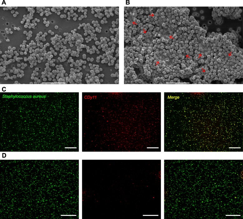
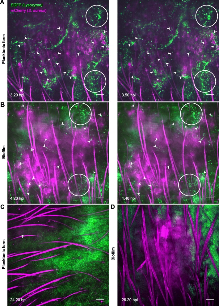
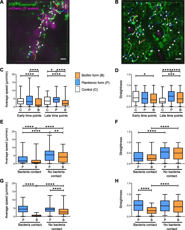
Results: We propose an in vivo transgenic mouse model, used in conjunction with intravital confocal microscopy to study the dynamics of host inflammatory responses to bacteria. Culture conditions were created to prepare calibrated inocula of fluorescent planktonic and biofilm forms of bacteria. A confocal imaging acquisition and analysis protocol was then drawn up to study the recruitment of innate immune cells in the skin of LysM-EGFP transgenic mice. Using the mouse ear pinna model, we showed that inflammatory responses to S. aureus can be quantified over time and that the dynamics of innate immune cells after injection of either the planktonic or biofilm form can be characterized. First results showed that the ability of phagocytic cells to infiltrate the injection site and their motility is not the same in planktonic and biofilm forms of bacteria despite the cells being considerably recruited in both cases.

Conclusion: We developed a mouse model of infection to compare the dynamics of the inflammatory responses to planktonic and biofilm bacteria at the tissue and cellular levels. The mouse ear pinna model is a powerful imaging system to analyse the mechanisms of biofilm tolerance to immune attacks.

Keywords: Biofilm; Inflammation; Intravital imaging; Mouse; Planktonic form; Staphylococcus aureus.


Conflict of interest statement

The authors declare that they have no competing interests.

Figures

Fig. 1
Fig. 1
Characterization of calibrated inocula of Staphylococcus aureus biofilm and planktonic cultures. a and b SEM micrographs of S. aureus LYO-S2 planktonic (a) and 24 h biofilm (b) inocula after passing through the 34G needle used for micro-injections. Red arrows in panel B indicate the biofilm extracellular matrix. Scale bar: 5 μm. c and d Fluorescence microscopy images of S. aureus biofilm (c) and planktonic (d) cultures stained with the green live cell fluorescent label SYTO9 and incubated with CDy11 red fluorescent probe. Scale bar: 50 μm
Fig. 2
Fig. 2
Micro-injection of calibrated inocula of Staphylococcus aureus in the mouse ear pinna. ac Reconstituted confocal images of the mouse ear pinna tissue showing the maximal projection intensities of the EGFP signal. LyM-EGFP transgenic mice were micro-injected with TS culture medium (a) or S. aureus mCherry-LYO-S2 in its planktonic (b) or biofilm (c) form at early (4–7 hpi) and late time points (after 24 hpi). The EGFP fluorescence (green) signal corresponds to phagocytic cells (neutrophils and macrophages). The yellow line indicates the ROI where the “Sum of EGFP fluorescence intensities” was measured. Scale bar: 2 mm. One representative experiment is shown for each group of mice from four independent experiments. d Ratio of the sum of EGFP fluorescence intensities to ROI area. Data are expressed as median and interquartile ranges for four mice per group
Fig. 3
Fig. 3
Dynamics of recruited EGFP+ cells in the mouse ear pinna after inoculation of Staphylococcus aureus. a and b Live confocal imaging after micro-injection of S. aureus mCherry-LYO-S2 in its planktonic (a) or biofilm (b) form in the ear pinna of LysM-EGFP transgenic mice at early time points. Innate immune cell recruitment towards planktonic bacteria and biofilms was observed between 3.20 to 3.50 hpi and 4.20 to 4.40 hpi, respectively. A progressive recruitment of EGFP+ innate immune cells was observed at the injection site with cell-bacteria contact areas (filled white arrowheads). White empty circles show cell accumulation over time for the planktonic or biofilm inoculum at early time points. *: autofluorescent hair (also in magenta). Scale bar: 100 μm. (c and d) Live confocal imaging at late time points after micro-injection of planktonic (c) or biofilm (d) bacteria, at 24.20 hpi and 26.20 hpi, respectively. Empty white arrowhead indicates the presence of remaining planktonic form after 24 h (low magenta signal) whereas biofilms were still easily detectable. Scale bar: 100 μm. a–d Images show average intensity projections of green (innate immune cells) and magenta (bacteria) fluorescence. One representative experiment is shown for each group of mice from three independent experiments
Fig. 4
Fig. 4
Motility of recruited EGFP+ cells in the mouse ear pinna after micro-injection of Staphylococcus aureus. a and b Illustration of immune cell tracking with Imaris software using the “Spots” tool to analyse the motility of recruited immune cells. The analysis was carried out in different zones of the injection site where cells were either in contact with visible bacteria (a) or not (b). Each cell is represented by a white sphere and its trajectory in the thickness of the tissue by a multicoloured line. Images shown were taken at 4.45 hpi (a) and 26 hpi (b). *: base of hair follicles. Scale bar: 50 μm. c–h Average speed and straightness of EGFP+ cells recruited to injection sites at early and late time points after inoculation of TS culture medium (control), planktonic bacteria (planktonic form) or biofilms (biofilm form). Data are expressed as median and interquartile ranges pooled from three different mice in three independent experiments for each group. Average speed (c) and straightness (d) of all cells (in contact with visible bacteria or not) in infected and control mice. Number of cells (N) analysed for each group at early and late time points, respectively: Control: N = 90 and 94 cells; Planktonic form: N = 315 and 433 cells; Biofilm form: N = 254 and 518 cells. Average speed (e and g) and straightness (f and h) of cells either in contact (bacteria contact) or not (no bacteria contact) with planktonic or biofilm bacteria at early (e and f) and late (g and h) time points. Number of cells (N) analysed at early time points that were in contact or not in contact with bacteria, respectively: Planktonic form: N = 157 and 158 cells; Biofilm form: N = 142 and 112 cells. Number of cells (N) analysed at late time points that were in contact or not in contact with bacteria, respectively: Planktonic form: N = 298 and 135 cells; Biofilm form: N = 98 and 420 cells

References

    1. Moormeier Derek E., Bayles Kenneth W. Staphylococcus aureusbiofilm: a complex developmental organism. Molecular Microbiology. 2017;104(3):365–376. doi: 10.1111/mmi.13634. - DOI - PMC - PubMed
    1. Ricciardi Benjamin F., Muthukrishnan Gowrishankar, Masters Elysia, Ninomiya Mark, Lee Charles C., Schwarz Edward M. Staphylococcus aureus Evasion of Host Immunity in the Setting of Prosthetic Joint Infection: Biofilm and Beyond. Current Reviews in Musculoskeletal Medicine. 2018;11(3):389–400. doi: 10.1007/s12178-018-9501-4. - DOI - PMC - PubMed
    1. Jamal M, Ahmad W, Andleeb S, Jalil F, Imran M, Nawaz MA, et al. Bacterial biofilm and associated infections. J Chin Med Assoc. 2018;81(1):7–11. doi: 10.1016/j.jcma.2017.07.012. - DOI - PubMed
    1. Yamada KJ, Kielian T. Biofilm-leukocyte cross-talk: impact on immune polarization and Immunometabolism. J Innate Immun. 2018;22:1–9. - PMC - PubMed
    1. Gries CM, Kielian T. Staphylococcal biofilms and immune polarization during prosthetic joint infection. J Am Acad Orthop Surg. 2017;25(Suppl 1):S20–S24. doi: 10.5435/JAAOS-D-16-00636. - DOI - PMC - PubMed

LinkOut - more resources