

Primary malignant melanoma of the vagina
- PMID: 31996380
- PMCID: PMC7021152
- DOI: 10.1136/bcr-2019-232200
Primary malignant melanoma of the vagina
Abstract
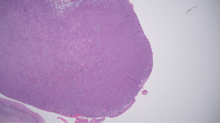
Primary malignant melanoma of the vagina is a rare gynaecological neoplasm with an aggressive course of disease. Although not many cases have been reported in the literature, its manifestations appear to be fairly consistent. The challenge comes in knowing how to approach this cancer clinically, since information about its staging and treatment is limited. In this report, we present a case of an 84-year-old postmenopausal woman in whom a suspicious vaginal lesion was discovered incidentally during a procedure. Wide local excision was carried out at a later date and histopathology confirmed a malignant melanoma of the vagina contained locally with no radiological finding of distant metastases. No additional treatment was given, and three monthly follow-ups were arranged for this patient. We review the literature and briefly discuss the epidemiology, treatment approaches, prognostic factors and expected outcomes of this rare disease.
Keywords: cancer intervention; gynecological cancer; obstetrics, gynaecology and fertility; pathology.
© BMJ Publishing Group Limited 2020. Re-use permitted under CC BY-NC. No commercial re-use. See rights and permissions. Published by BMJ.
Conflict of interest statement
Competing interests: None declared.
Figures


References
-
- Chang AE, Karnell LH, Menck HR. The National cancer data base report on cutaneous and noncutaneous melanoma: a summary of 84,836 cases from the past decade. the American College of surgeons Commission on cancer and the American cancer Society. Cancer 1998;83:1664–78. 10.1002/(sici)1097-0142(19981015)83:8<1664::aid-cncr23>3.0.co;2-g - DOI - PubMed
Publication types
MeSH terms
LinkOut - more resources
Full Text Sources
Medical