

Rare case of acute on chronic hepatic failure in a patient with multiple myeloma-associated amyloidosis
- PMID: 31996385
- PMCID: PMC7021185
- DOI: 10.1136/bcr-2019-231563
Rare case of acute on chronic hepatic failure in a patient with multiple myeloma-associated amyloidosis
Abstract
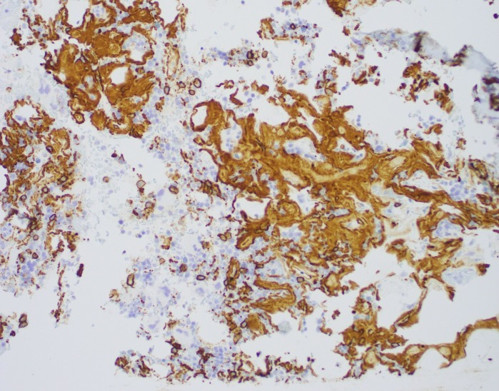
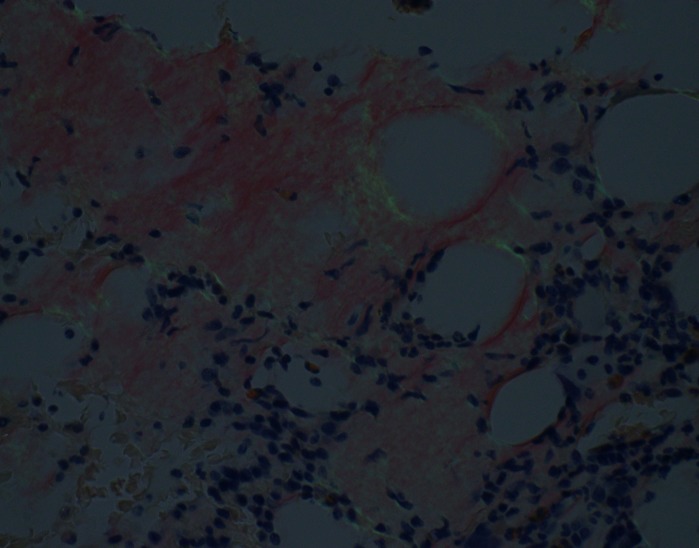
Amyloidosis is the extracellular deposition of unique protein fibrils in different tissue organs. It is most commonly associated with B-cell malignancy such as multiple myeloma or Waldenstrom macroglobulinaemia. It involves the liver, heart, kidney, peripheral nerves and soft tissues. Liver however is affected, but clinically apparent disease is very rare. Hepatomegaly and mild elevation of alkaline phosphatase is the most common presentation in patients with liver involvement. Acute hepatic failure is a rare presentation with myeloma-induced amyloidosis. The diagnosis can be difficult requiring biopsy or sometimes special staining of the tissue. Management is still very challenging.
Keywords: gastroenterology; liver disease; oncology.
© BMJ Publishing Group Limited 2020. No commercial re-use. See rights and permissions. Published by BMJ.
Conflict of interest statement
Competing interests: None declared.
Figures


Similar articles
-
Myeloma associated amyloidosis presenting as subacute liver failure.Postgrad Med J. 2006 Jul;82(969):e13. doi: 10.1136/pgmj.2006.044883. Postgrad Med J. 2006. PMID: 16822912 Free PMC article.
-
Multiple Myeloma Associated Intestinal Amyloidosis: Intestinal Pseudoobstruction Falsely Considered as an Ascites.Rev Recent Clin Trials. 2018 Jan 31;13(1):79-81. doi: 10.2174/0929867324666170830114914. Rev Recent Clin Trials. 2018. PMID: 28875827
-
Acute liver failure due to primary amyloidosis in a nephrotic syndrome: a swiftly progressive course.BMJ Case Rep. 2016 Mar 10;2016:bcr2016214392. doi: 10.1136/bcr-2016-214392. BMJ Case Rep. 2016. PMID: 26965175 Free PMC article.
-
Hepatic failure in a case of multiple myeloma-associated amyloidosis (kappa-AL).J Gastroenterol. 1995 Jun;30(3):393-7. doi: 10.1007/BF02347517. J Gastroenterol. 1995. PMID: 7647907 Review.
-
Primary systemic amyloidosis initially presenting with digestive symptoms: a case report and review of the literature.Diagn Pathol. 2015 Sep 21;10:174. doi: 10.1186/s13000-015-0407-9. Diagn Pathol. 2015. PMID: 26390868 Free PMC article. Review.
Cited by
-
miRNAs and Multiple Myeloma: Focus on the Pathogenesis, Prognosis, and Drug Resistance.Technol Cancer Res Treat. 2023 Jan-Dec;22:15330338231202391. doi: 10.1177/15330338231202391. Technol Cancer Res Treat. 2023. PMID: 37728167 Free PMC article. Review.
References
-
- Serra L, Poppi MC, Criscuolo M, et al. . Primary systemic amyloidosis with giant hepatomegaly and portal hypertension: a case report and a review of the literature. Ital J Gastroenterol 1993;25:435–8. - PubMed
-
- Bujanda L, Beguiristain A, Alberdi F, et al. . Spontaneous rupture of the liver in amyloidosis. Am J Gastroenterol 1997;92:1385–6. - PubMed
Publication types
MeSH terms
LinkOut - more resources
Full Text Sources
Medical