. 2020 Mar 31;94(8):e01997-19.
doi: 10.1128/JVI.01997-19. Print 2020 Mar 31.

Characterization of Mollivirus kamchatka, the First Modern Representative of the Proposed Molliviridae Family of Giant Viruses

Affiliations

Characterization of Mollivirus kamchatka, the First Modern Representative of the Proposed Molliviridae Family of Giant Viruses

Eugene Christo-Foroux et al. J Virol. .

Abstract

Microbes trapped in permanently frozen paleosoils (permafrost) are the focus of increasing research in the context of global warming. Our previous investigations led to the discovery and reactivation of two Acanthamoeba-infecting giant viruses, Mollivirus sibericum and Pithovirus sibericum, from a 30,000-year old permafrost layer. While several modern pithovirus strains have since been isolated, no contemporary mollivirus relative was found. We now describe Mollivirus kamchatka, a close relative to M. sibericum, isolated from surface soil sampled on the bank of the Kronotsky River in Kamchatka, Russian Federation. This discovery confirms that molliviruses have not gone extinct and are at least present in a distant subarctic continental location. This modern isolate exhibits a nucleocytoplasmic replication cycle identical to that of M. sibericum Its spherical particle (0.6 μm in diameter) encloses a 648-kb GC-rich double-stranded DNA genome coding for 480 proteins, of which 61% are unique to these two molliviruses. The 461 homologous proteins are highly conserved (92% identical residues, on average), despite the presumed stasis of M. sibericum for the last 30,000 years. Selection pressure analyses show that most of these proteins contribute to virus fitness. The comparison of these first two molliviruses clarify their evolutionary relationship with the pandoraviruses, supporting their provisional classification in a distinct family, the Molliviridae, pending the eventual discovery of intermediary missing links better demonstrating their common ancestry.IMPORTANCE Virology has long been viewed through the prism of human, cattle, or plant diseases, leading to a largely incomplete picture of the viral world. The serendipitous discovery of the first giant virus visible under a light microscope (i.e., >0.3 μm in diameter), mimivirus, opened a new era of environmental virology, now incorporating protozoan-infecting viruses. Planet-wide isolation studies and metagenome analyses have shown the presence of giant viruses in most terrestrial and aquatic environments, including upper Pleistocene frozen soils. Those systematic surveys have led authors to propose several new distinct families, including the Mimiviridae, Marseilleviridae, Faustoviridae, Pandoraviridae, and Pithoviridae We now propose to introduce one additional family, the Molliviridae, following the description of M. kamchatka, the first modern relative of M. sibericum, previously isolated from 30,000-year-old arctic permafrost.

Keywords: Acanthamoeba; Arctic; Kamchatka; NCLDV; comparative genomics; paleovirology.


Figures

FIG 1
FIG 1
Phylogeny of DNA polymerase B of large and giant dsDNA viruses. This neighbor-joining tree was computed (JTT substitution model, 100 resamplings) on 397 amino acid positions from an alignment of 42 sequences computed by the MAFFT program (29). Branches with bootstrap values of <60% were collapsed.
FIG 2
FIG 2
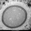
Ultra-thin-section TEM image of a newly synthesized M. kamchatka particle in the cell cytoplasm at 7 h postinfection. The structure of the mature particles appears to be identical to that of M. sibericum mature particles.
FIG 3
FIG 3
Ultra-thin-section TEM image of an A. castellanii cell at 7 to 10 h postinfection by M. kamchatka. (A) A viral factory exhibiting fibrils (F), a nascent viral particle (V), and surrounding mitochondria (M). Fragments of the ruptured nuclear membrane are visible as dark bead strings. (B) Details of a nuclear membrane rupture through which fibrils synthesized in the nucleus (N) are shed into the cytoplasm (C).
FIG 4
FIG 4
Distribution of the best-matching nonredundant homologs of M. kamchatka predicted proteins. Best-matching homologous proteins were identified using the BLASTp program (E value, <10−5) against the nonredundant (NR) database (15) (after excluding M. sibericum). Green shades are used for eukaryotes, and red shades are used for viruses.
FIG 5
FIG 5
Eventual gene transfers from a pandoravirus to M. kamchatka. Both phylogenetic trees were computed from the global alignments of orthologous protein sequences using MAFFT (29). IQtree (48) was used to determine the optimal substitution model (options, -m TEST and –bb 1000). (A) Results for the mk_165 protein (no predicted function). The corresponding long branch suggests its accelerated divergence since an ancient acquisition from a pandoravirus. (B) Results for the predicted methyltransferase mk_92. The long branch leading to the P. dulcis homolog might alternatively be interpreted as a nonorthologous replacement of the ancestral pandoravirus version of the gene.
FIG 6
FIG 6
Genomic features of strain-specific genes encoding ORFans. (A) Codon adaption index (CAI); (B) G+C content; (C) protein length. The box plots show the median and the 25th and 75th percentiles. P values were calculated using the Wilcoxon test.
FIG 7
FIG 7
Selection pressure among different classes of genes. Values of ω (i.e., dN/dS) were computed from the alignments of homologous coding regions in M. kamchatka and M. sibericum. (A) Distribution of calculated ω values (n = 397). (B) Box plots of the ω ratio among ORFan genes (n = 243) and non-ORFan genes (n = 154). The box plots show the median and the 25th and 75th percentiles. All P values were calculated using the Wilcoxon test.
FIG 8
FIG 8
Comparison of the mollivirus and pandoravirus core gene contents. (A) The distribution of the protein clusters shared by all pandoraviruses (black), by the two Molliviruses (pink), and by both virus groups (super core genes) (blue). (B) Box plot of ω values calculated from the alignment of mollivirus core genes (pink) and super core genes (blue). The box plots show the median and the 25th and 75th percentiles.
FIG 9
FIG 9
Distribution of different classes of genes along mollivirus genomes. (A) Variation of the gene density computed by use of the ggpplot2 geom_density function (49). The distribution of super core genes (n = 64, in green) is strongly biased toward the right half of the genome, in contrast to the genes with best-matching homologs in A. castellanii (in the nonredundant database, excluding mollivirus), which are more evenly distributed (in pink) (n = 55 and n = 51 for M. sibericum and M. kamchatka, respectively). M. sibericum-specific ORFan-encoding genes (in blue) also exhibit a nonuniform distribution toward the left half of the genome. (B) Cumulative distribution of the above-described classes of genes using the same color code used in panel A.
FIG 10
FIG 10
Distribution of single-copy versus multiple-copy genes along mollivirus genomes. Single-copy genes (in blue) in both strains are evenly distributed, in contrast to genes with paralogs (pink), which cluster in the left half of the genomes. (A) M. sibericum (n = 48); (B) M. kamchatka (n = 46).

References

    1. La Scola B, Audic S, Robert C, Jungang L, de Lamballerie X, Drancourt M, Birtles R, Claverie JM, Raoult D. 2003. A giant virus in amoebae. Science 299:2033. doi:10.1126/science.1081867. - DOI - PubMed
    1. Raoult D, Audic S, Robert C, Abergel C, Renesto P, Ogata H, La Scola B, Suzan M, Claverie JM. 2004. The 1.2-megabase genome sequence of mimivirus. Science 306:1344–1350. doi:10.1126/science.1101485. - DOI - PubMed
    1. Claverie JM, Grzela R, Lartigue A, Bernadac A, Nitsche S, Vacelet J, Ogata H, Abergel C. 2009. Mimivirus and Mimiviridae: giant viruses with an increasing number of potential hosts, including corals and sponges. J Invertebr Pathol 101:172–180. doi:10.1016/j.jip.2009.03.011. - DOI - PubMed
    1. Abergel C, Legendre M, Claverie JM. 2015. The rapidly expanding universe of giant viruses: Mimivirus, Pandoravirus, Pithovirus and Mollivirus. FEMS Microbiol Rev 39:779–796. doi:10.1093/femsre/fuv037. - DOI - PubMed
    1. Lefkowitz EJ, Dempsey DM, Hendrickson RC, Orton RJ, Siddell SG, Smith DB. 2018. Virus taxonomy: the database of the International Committee on Taxonomy of Viruses (ICTV). Nucleic Acids Res 46:D708–D717. doi:10.1093/nar/gkx932. - DOI - PMC - PubMed

Publication types

MeSH terms