. 2020 Mar 25;40(13):2633-2643.
doi: 10.1523/JNEUROSCI.2374-19.2020. Epub 2020 Jan 29.

Cortical and Subcortical Effects of Transcutaneous Spinal Cord Stimulation in Humans with Tetraplegia

Affiliations

Cortical and Subcortical Effects of Transcutaneous Spinal Cord Stimulation in Humans with Tetraplegia

Francisco D Benavides et al. J Neurosci. .

Abstract

An increasing number of studies supports the view that transcutaneous electrical stimulation of the spinal cord (TESS) promotes functional recovery in humans with spinal cord injury (SCI). However, the neural mechanisms contributing to these effects remain poorly understood. Here we examined motor-evoked potentials in arm muscles elicited by cortical and subcortical stimulation of corticospinal axons before and after 20 min of TESS (30 Hz pulses with a 5 kHz carrier frequency) and sham-TESS applied between C5 and C6 spinous processes in males and females with and without chronic incomplete cervical SCI. The amplitude of subcortical, but not cortical, motor-evoked potentials increased in proximal and distal arm muscles for 75 min after TESS, but not sham-TESS, in control subjects and SCI participants, suggesting a subcortical origin for these effects. Intracortical inhibition, elicited by paired stimuli, increased after TESS in both groups. When TESS was applied without the 5 kHz carrier frequency both subcortical and cortical motor-evoked potentials were facilitated without changing intracortical inhibition, suggesting that the 5 kHz carrier frequency contributed to the cortical inhibitory effects. Hand and arm function improved largely when TESS was used with, compared with without, the 5 kHz carrier frequency. These novel observations demonstrate that TESS influences cortical and spinal networks, having an excitatory effect at the spinal level and an inhibitory effect at the cortical level. We hypothesized that these parallel effects contribute to further the recovery of limb function following SCI.SIGNIFICANCE STATEMENT Accumulating evidence supports the view that transcutaneous electrical stimulation of the spinal cord (TESS) promotes recovery of function in humans with spinal cord injury (SCI). Here, we show that a single session of TESS over the cervical spinal cord in individuals with incomplete chronic cervical SCI influenced in parallel the excitability cortical and spinal networks, having an excitatory effect at the spinal level and an inhibitory effect at the cortical level. Importantly, these parallel physiological effects had an impact on the magnitude of improvements in voluntary motor output.

Keywords: corticospinal; intracortical inhibition; neurophysiology; neuroplasticity; spinal cord injury; spinal networks.


Figures

Figure 1.
Figure 1.
Experimental setup. A, Participants were comfortably seated in a customized chair during TESS or sham-TESS for 20 min. TESS was delivered using a surface electrode on the back of the neck between C5 and C6 spinous processes segments (cathode) and a surface electrode in each anterior crest of the hip bone (anode) using a custom-made five-channel stimulator (BioStim5). Electrophysiological and behavioral outcomes were tested Pre, immediately after, and 15, 30, 45, 60, and 75 min after the end of the stimulation or sham stimulation period. B, Schematic representation of the type of current used during TESS. We used five biphasic pulses at 5 kHz with each biphasic pulse lasting for 200 μs. The middle scheme shows the blocks of five biphasic pulses passed at a 30 Hz frequency. The bottom part of the schematic shows the number of pulses delivered in 1 s.
Figure 2.
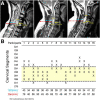
Figure 2.
Structural MRI. A, Three sagittal slices from representative participants with cervical SCI. The blue arrows show the distance from the canal toward the beginning of the SC space. The red arrows show the distance from the spinal canal toward the surface of the skin, including the cutaneous layer and fat tissue underneath. The yellow dotted lines show the top and bottom borders of the injury, and arrows point to the C5–C6 intervertebral space. Individual distances and segmental distributions of the injuries are shown in B.
Figure 3.
Figure 3.
CMEPs. A, B, Averaged CMEP traces in the biceps brachii muscle in a representative control subject (A) and a participant with SCI (B) before and after TESS and sham-TESS. Each waveform represents the average of 10 CMEPs. Graphs show group (control subjects, n = 15; SCI, n = 17) and individual data. The ordinate shows the CMEP amplitude as a percentage of the CMEP at baseline (percentage of baseline) in control subjects and SCI participants before and after TESS (control subjects, green circles; SCI participants, orange circles) and sham-TESS (control subjects and SCI participants, black circles). The abscissa shows the time of measurements (Pre, and 15, 30, 45, 60, and 75 min after each protocol). Note that in graphs showing data from individual participant postmeasurement data showed the average from 15 to 75 min. Error bars indicate SDs. *p < 0.05.
Figure 4.
Figure 4.
MEPs. A, B, Averaged MEP traces elicited by TMS in the biceps brachii muscle in a representative participant before and after each protocol. Each waveform represents the average of 20 MEPs. Graphs show group (control subjects, n = 15; SCI participants, n = 17) and individual data. The ordinate shows the MEP amplitude as a percentage of the MEP at baseline (percentage of baseline) in control subjects and SCI participants before and after TESS (control subjects, green circles; SCI participants, orange circles) and sham-TESS (control subjects and SCI participants, black circles). The abscissa shows the time of measurements (Pre, and 15, 30, 45, 60, and 75 min after each protocol). Error bars indicate SDs. *p < 0.05.
Figure 5.
Figure 5.
SICI. A, B, Averaged traces showing SICI measurements in the biceps brachii muscle in a representative control subject (A) and SCI participant (B) before and after TESS. Each waveform represents the average of 15 test MEPs (black traces) and 15 conditioned MEPs (gray traces). Arrows at the beginning of each trace indicate the TS and CS used during testing. Graphs show group data (control subjects, n = 15; SCI participants, n = 17) and individual data. The ordinate shows the conditioned MEP as a percentage of the test MEP at baseline (percentage of test response) in control subjects and SCI participants before and after TESS (control subjects, green circles; SCI participants, orange circles). The abscissa shows the time of measurements (Pre, and 15, 30, 45, 60, and 75 min after each protocol). In graphs showing data from individual participants, postmeasurements show the average from 15 to 75 min. Error bars indicate SDs. *p < 0.05.
Figure 6.
Figure 6.
Effect of different currents. A–B, Graphs show group data (A. control subjects, n = 8; B. SCI participants, n = 8) for CMEPs MEPs and SICI before and after the TESS, sham-TESS, and TESS applied without the 5 kHz carrier frequency (TESS w/o 5 kHz) in control subjects and SCI participants, respectively. The ordinate shows CMEP and MEP amplitude as a percentage of CMEPs and MEPs at baseline (percentage of baseline) and SICI as a percentage of SICI at baseline (percentage of baseline) in control subjects and SCI participants before and after TESS (light gray circles), sham-TESS (black circles), and TESS without 5 kHz (dark gray circles). The abscissa shows the time of measurements (Pre, and 15, 30, 45, 60, and 75 min after each protocol). Error bars indicate SDs. *p < 0.05.
Figure 7.
Figure 7.
Functional outcomes. A, B, Images showing the three subcomponents of the GRASSP (coint, key, and water bottle tests, respectively) tested in SCI participants (n = 10) before and ∼75 min after sham-TESS, TESS w/o 5 kHz, and TESS. Graphs show individual (left side, A) and group (right side, B). The ordinate shows the time to complete each task as a percentage of the time needed at baseline (percentage of baseline) before and after TESS (light gray circles), sham-TESS (black circles), and TESS without 5 kHz (dark gray circles). The abscissa shows the subcomponents of the GRASSP tested (ccoint, key, and water bottle tests). Error bars indicate SDs. *p < 0.05.

References

    1. Alam M, Garcia-Alias G, Jin B, Keyes J, Zhong H, Roy RR, Gerasimenko Y, Lu DC, Edgerton VR (2017) Electrical neuromodulation of the cervical spinal cord facilitates forelimb skilled function recovery in spinal cord injured rats. Exp Neurol 291:141–150. 10.1016/j.expneurol.2017.02.006 - DOI - PMC - PubMed
    1. Awosika OO, Sandrini M, Volochayev R, Thompson RM, Fishman N, Wu T, Floeter MK, Hallett M, Cohen LG (2019) Transcutaneous spinal direct current stimulation improves locomotor learning in healthy humans. Brain Stimul 12:628–634. 10.1016/j.brs.2019.01.017 - DOI - PMC - PubMed
    1. Berardelli A, Inghilleri M, Cruccu G, Manfredi M (1991) Corticospinal potentials after electrical and magnetic stimulation in man. Electroencephalogr Clin Neurophysiol Suppl 43:147–154. - PubMed
    1. Bertolasi L, Priori A, Tinazzi M, Bertasi V, Rothwell JC (1998) Inhibitory action of forearm flexor muscle afferents on corticospinal outputs to antagonist muscles in humans. J Physiol 511:947–956. 10.1111/j.1469-7793.1998.947bg.x - DOI - PMC - PubMed
    1. Bunday KL, Perez MA (2012) Motor recovery after spinal cord injury enhanced by strengthening corticospinal synaptic transmission. Curr Biol 22:2355–2361. 10.1016/j.cub.2012.10.046 - DOI - PMC - PubMed

Publication types

MeSH terms