Review
. 2020 Jan 30;5(2):e132964.
doi: 10.1172/jci.insight.132964.

Regulation of tissue iron homeostasis: the macrophage "ferrostat"

Affiliations
Review

Regulation of tissue iron homeostasis: the macrophage "ferrostat"

Nathan C Winn et al. JCI Insight. .

Abstract

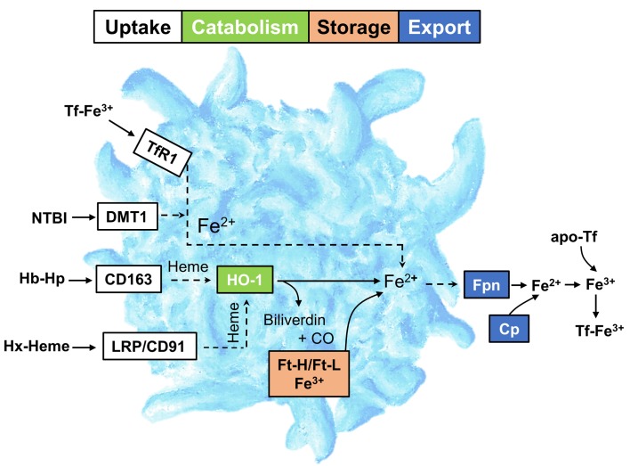
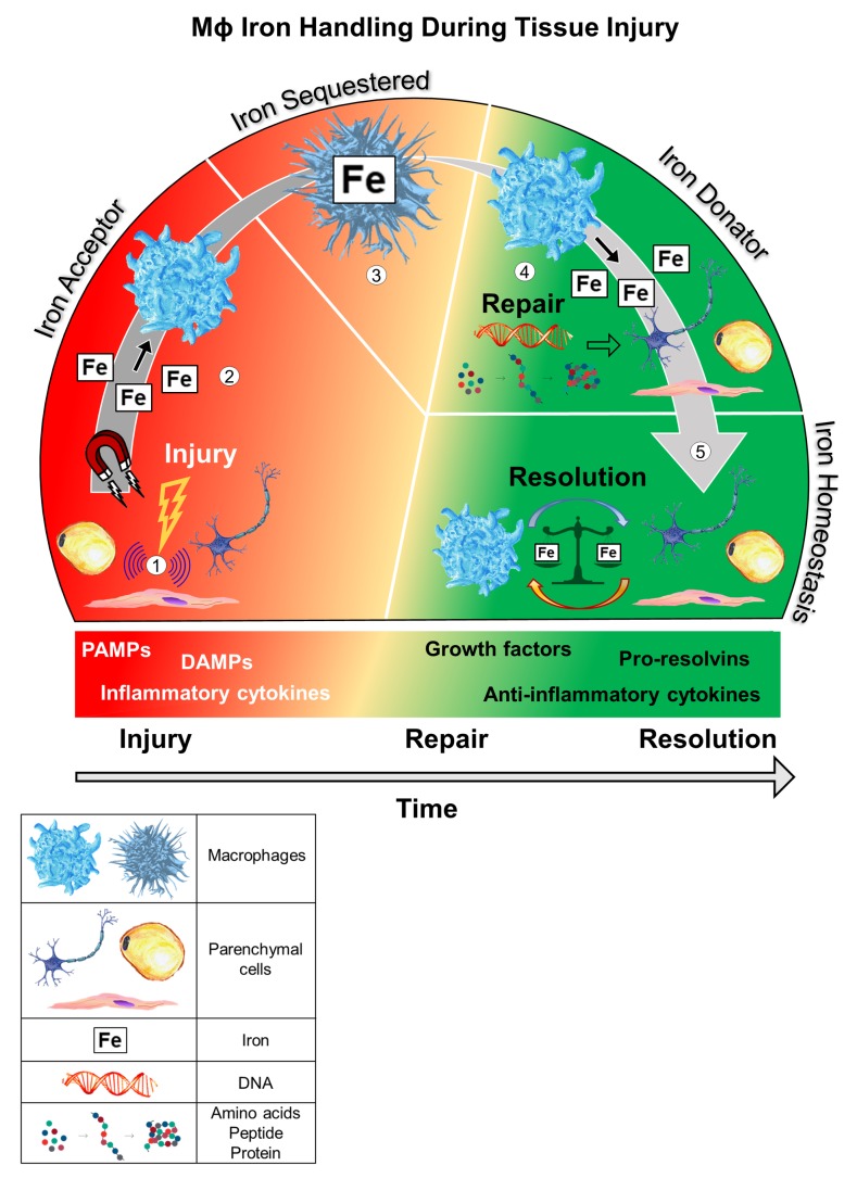
Iron is an essential element for multiple fundamental biological processes required for life; yet iron overload can be cytotoxic. Consequently, iron concentrations at the cellular and tissue level must be exquisitely governed by mechanisms that complement and fine-tune systemic control. It is well appreciated that macrophages are vital for systemic iron homeostasis, supplying or sequestering iron as needed for erythropoiesis or bacteriostasis, respectively. Indeed, recycling of iron through erythrophagocytosis by splenic macrophages is a major contributor to systemic iron homeostasis. However, accumulating evidence suggests that tissue-resident macrophages regulate local iron availability and modulate the tissue microenvironment, contributing to cellular and tissue function. Here, we summarize the significance of tissue-specific regulation of iron availability and highlight how resident macrophages are critical for this process. This tissue-dependent regulation has broad implications for understanding both resident macrophage function and tissue iron homeostasis in health and disease.


Conflict of interest statement

Conflict of interest: The authors have declared that no conflict of interest exists.

Figures

Figure 1
Figure 1. Overview of Mɸ iron metabolism.
The uptake of Tf-bound Fe3+ is mediated by TfR1. CD163 and LRP1 (also known as CD91) mediate the uptake of Hb-Hp and Hx-heme, respectively. Intracellular heme-Fe (extracted from Hb-Hp and Hx-heme) is catabolized by HO-1 for incorporation into the cellular ferritin pool or trafficked into the mitochondria. Non-Tf-bound iron (NTBI) is imported via DMT1. Iron export by Fpn is facilitated by Cp, which oxidizes Fe2+ to Fe3+ to allow sequestration by apo-Tf. Tf, transferrin; TfR1, transferrin receptor 1; LRP/CD91, LDL-related receptor 1/CD91; DMT1, divalent metal transporter 1; Hb-Hp, hemoglobin-haptoglobin; Hx-heme, hemopexin-heme; HO-1, heme oxygenase 1; Ft-H, ferritin heavy chain 1; Ft-L, ferritin light chain; Fpn, ferroportin; Cp, ceruloplasmin.
Figure 2
Figure 2. Proposed model by which resident iron-handling Mɸs regulate tissue homeostasis during an insult.
(1) Parenchymal cell injury initiates intracellular transduction signals that propagate transcriptional and nontranscriptional stress signals. (2) Local tissue-resident Mɸs are recruited to the injured site. Since injured cells are more susceptible to iron-induced oxidative damage, Mɸs sequester extracellular iron to decrease iron uptake in the injured parenchymal cell. LIP has been reported to increase in response to cell injury, which may promote iron efflux and Mɸ uptake. (3) A latency period follows, in which the Mɸ retains sequestered iron, allowing for parenchymal cell restoration. This phase of Mɸ iron retention may be aided by release of inflammatory cytokines and/or IFN responses that favor an iron-loaded Mɸ (i.e., suppression of Fpn-mediated iron export). Parenchymal cell repair requires iron for processes such as DNA synthesis. This utilization of parenchymal intracellular iron depletes ferritin stores. (4) The Mɸ mobilizes and relinquishes iron for parenchymal cell repair. (5) An undefined termination signal communicates cell resolution and the Mɸ regresses from the site, completing the homeostatic circuit and maintaining local iron balance.

References

    1. Musallam KM, Taher AT. Iron deficiency beyond erythropoiesis: should we be concerned? Curr Med Res Opin. 2018;34(1):81–93. doi: 10.1080/03007995.2017.1394833. - DOI - PubMed
    1. von Haehling S, Jankowska EA, van Veldhuisen DJ, Ponikowski P, Anker SD. Iron deficiency and cardiovascular disease. Nat Rev Cardiol. 2015;12(11):659–669. doi: 10.1038/nrcardio.2015.109. - DOI - PubMed
    1. Fernandez-Real JM, Manco M. Effects of iron overload on chronic metabolic diseases. Lancet Diabetes Endocrinol. 2014;2(6):513–526. doi: 10.1016/S2213-8587(13)70174-8. - DOI - PubMed
    1. Ward RJ, Zucca FA, Duyn JH, Crichton RR, Zecca L. The role of iron in brain ageing and neurodegenerative disorders. Lancet Neurol. 2014;13(10):1045–1060. doi: 10.1016/S1474-4422(14)70117-6. - DOI - PMC - PubMed
    1. Pantopoulos K, Porwal SK, Tartakoff A, Devireddy L. Mechanisms of mammalian iron homeostasis. Biochemistry. 2012;51(29):5705–5724. doi: 10.1021/bi300752r. - DOI - PMC - PubMed