. 2020 Feb;578(7793):166-171.
doi: 10.1038/s41586-020-1952-2. Epub 2020 Jan 29.

PIK3CA variants selectively initiate brain hyperactivity during gliomagenesis

Affiliations

PIK3CA variants selectively initiate brain hyperactivity during gliomagenesis

Kwanha Yu et al. Nature. 2020 Feb.

Abstract

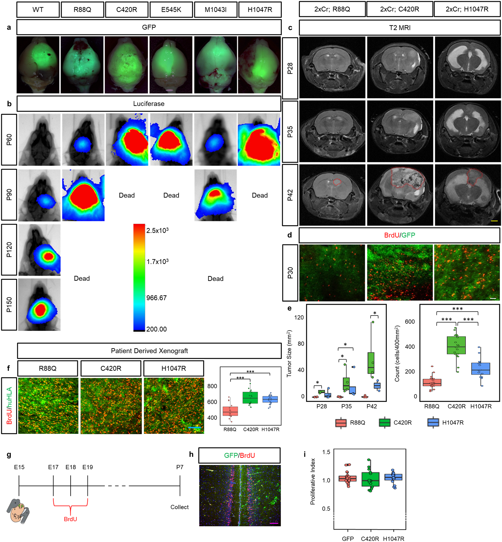
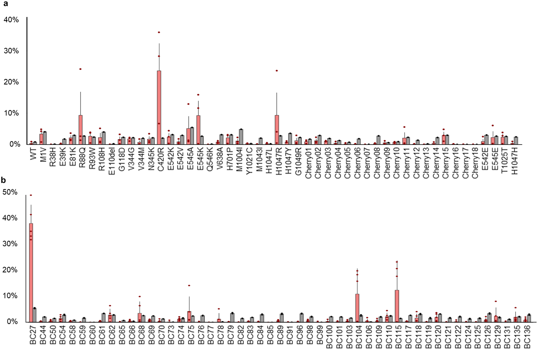
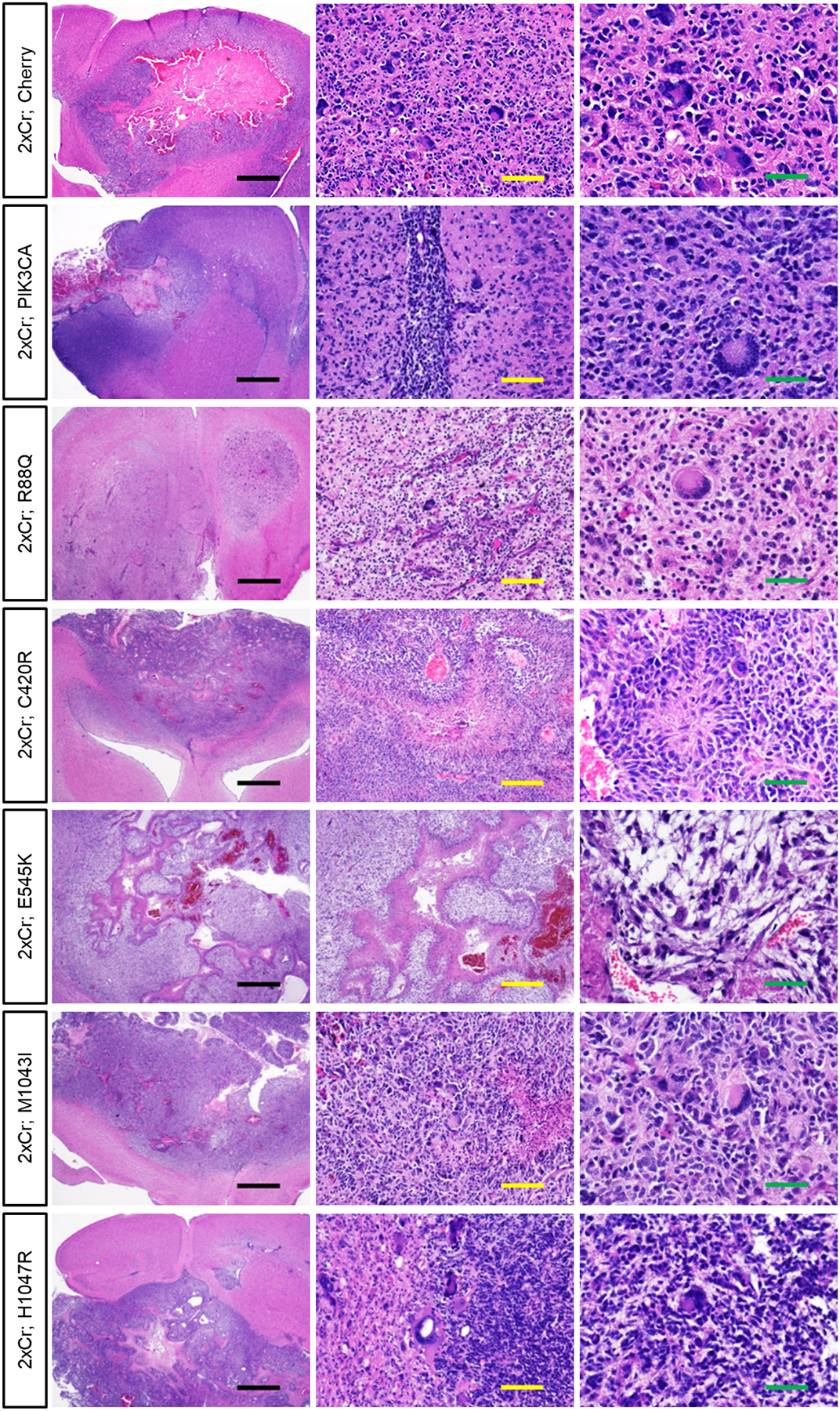
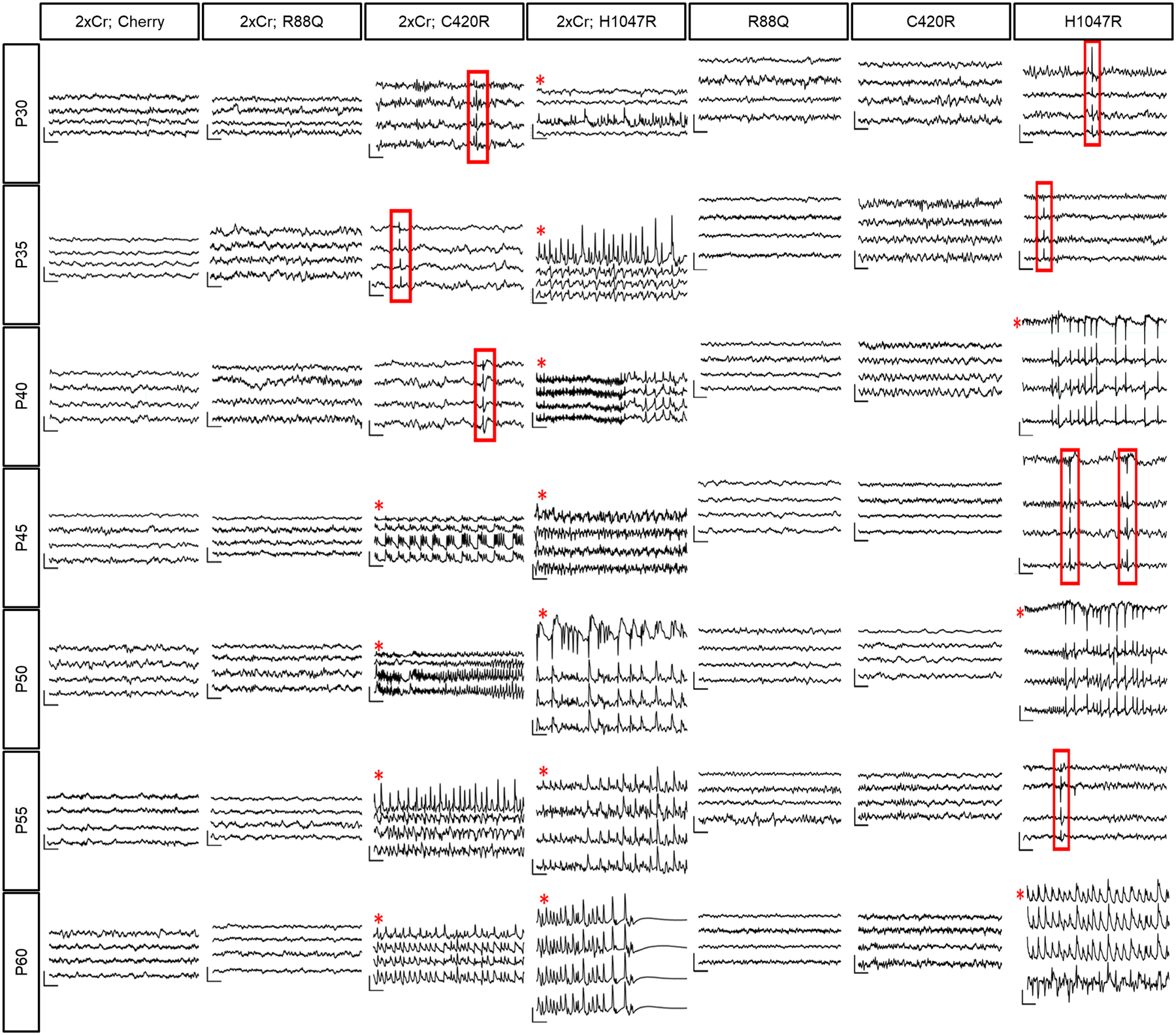
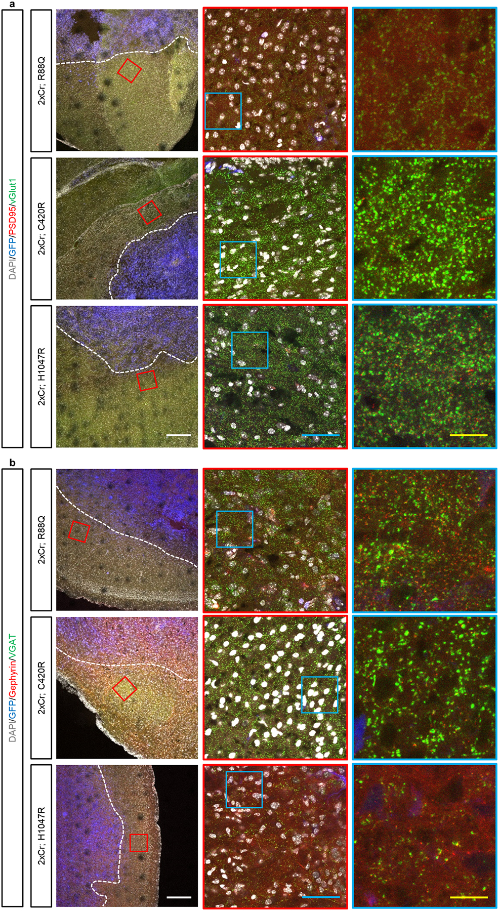
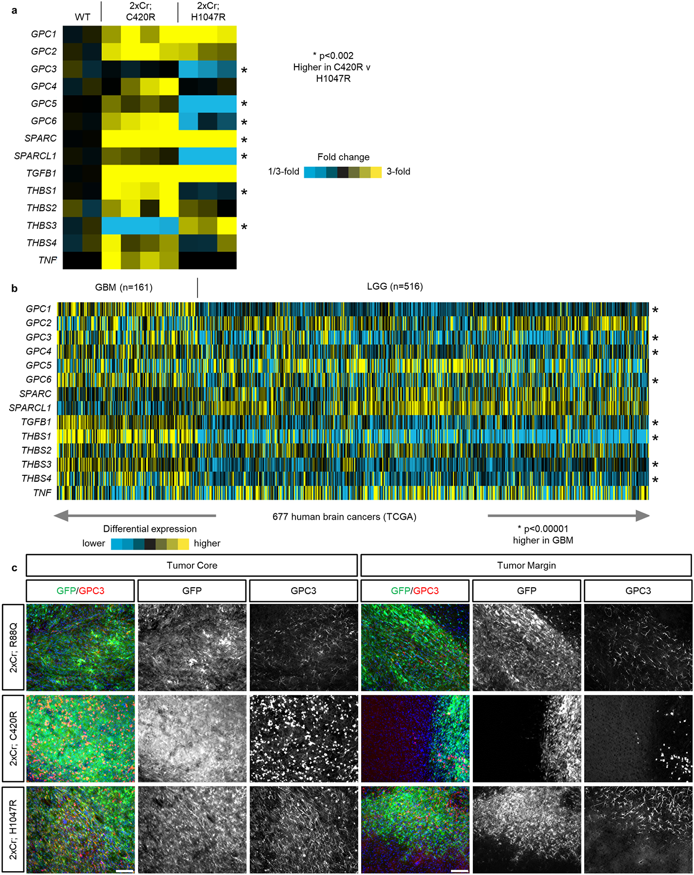
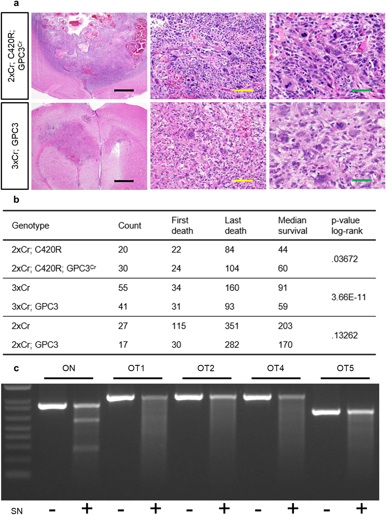
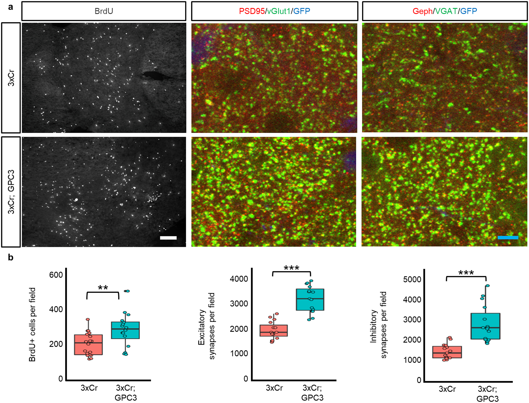
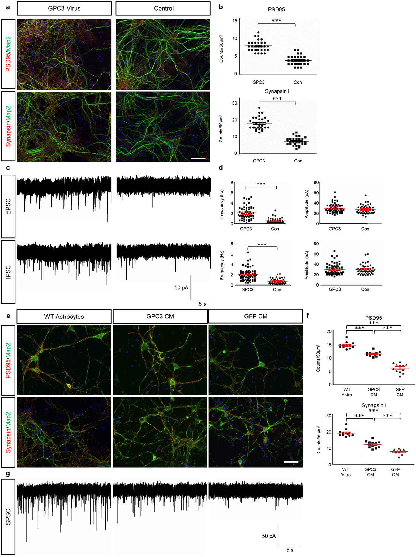
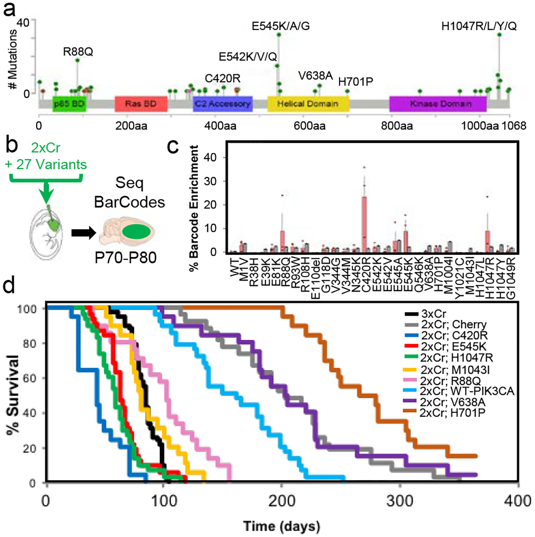
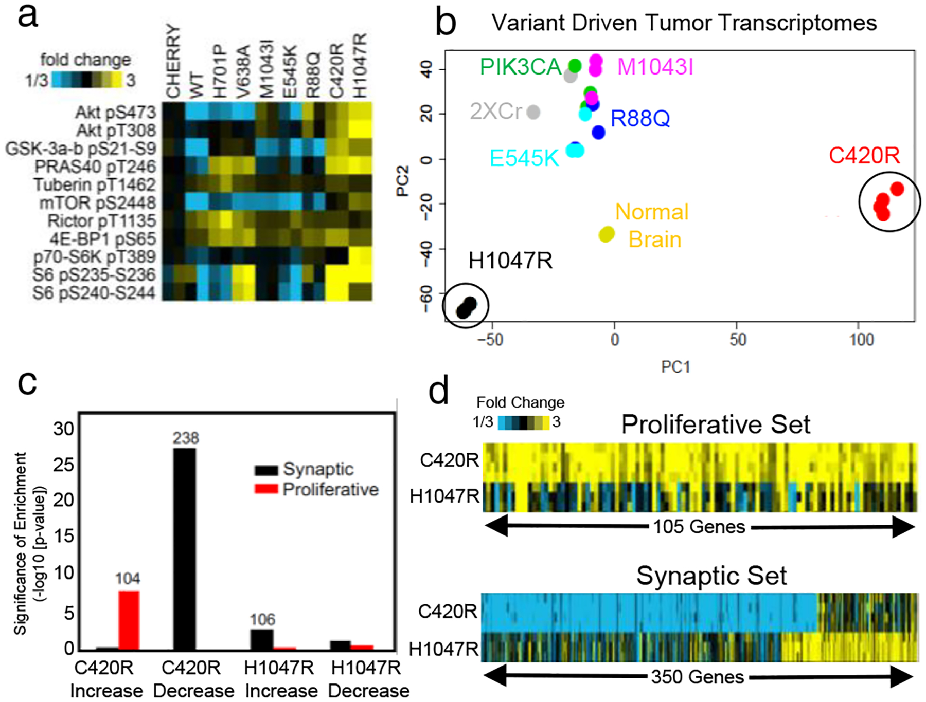
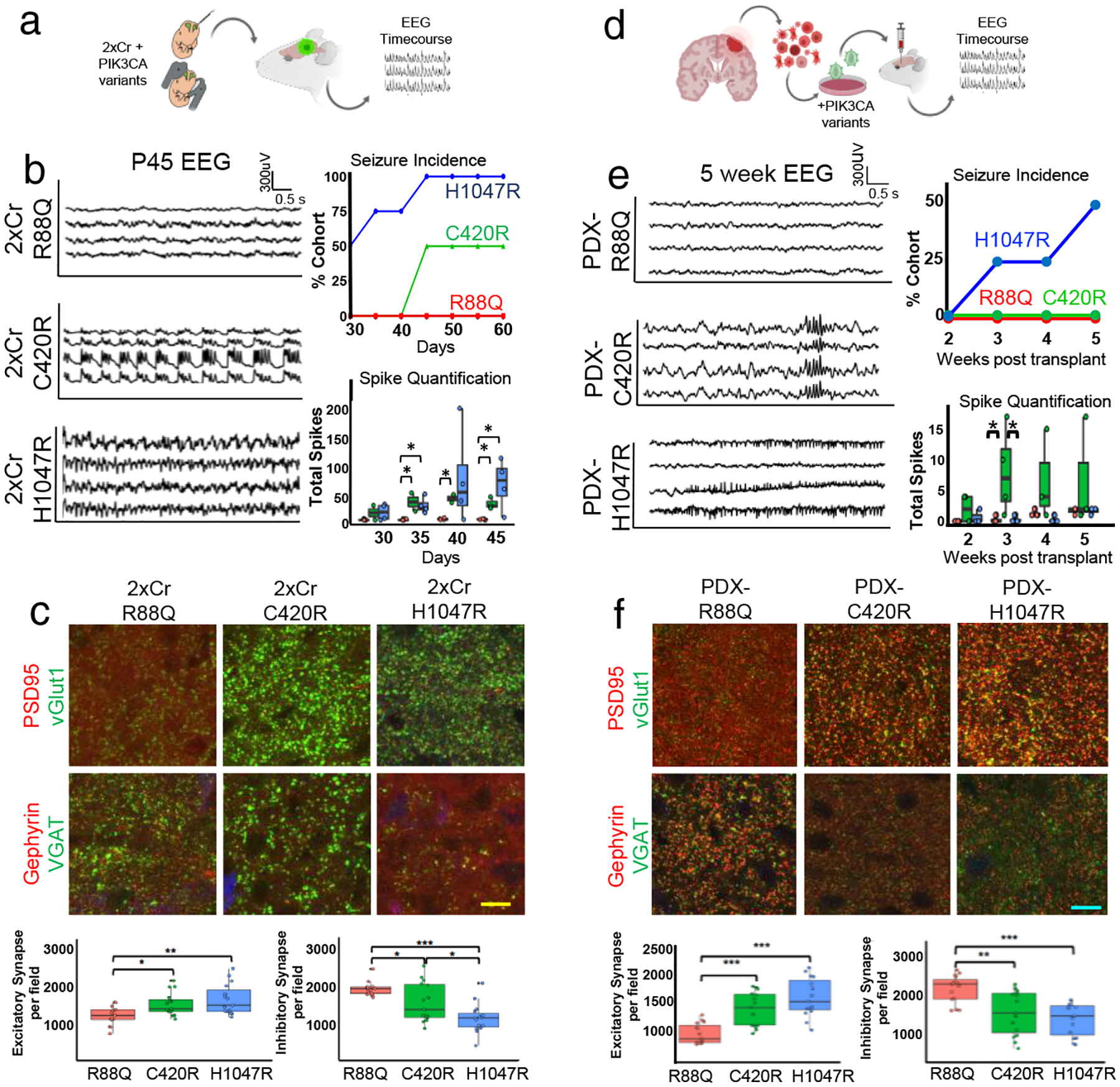
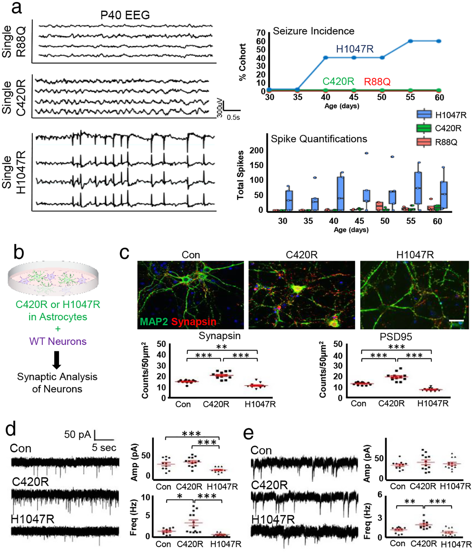
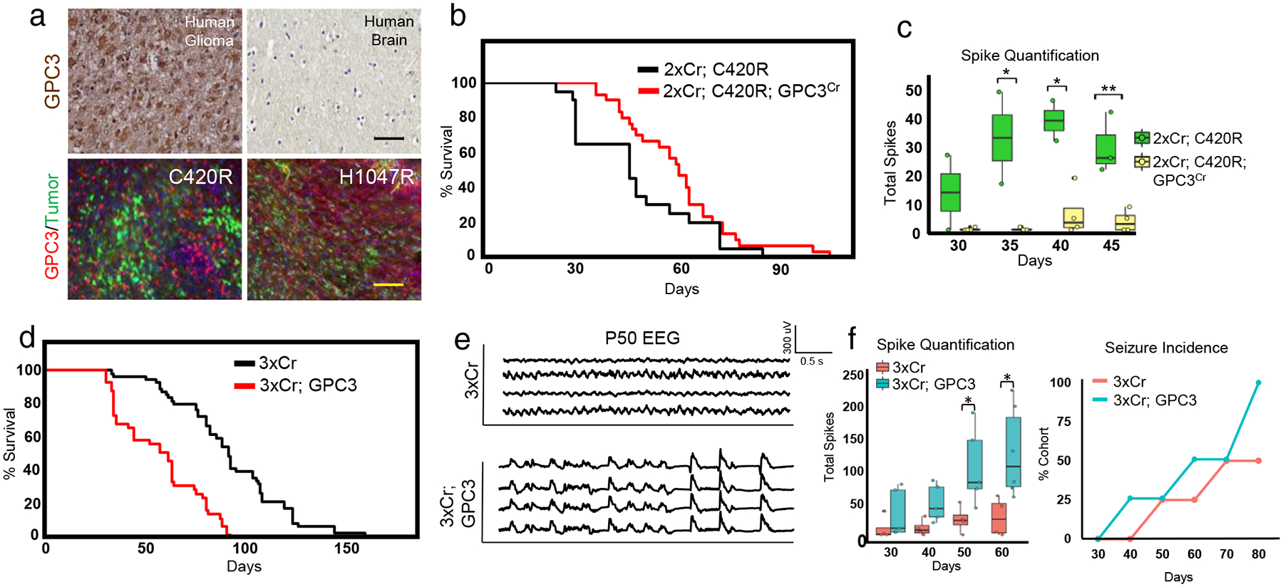
Glioblastoma is a universally lethal form of brain cancer that exhibits an array of pathophysiological phenotypes, many of which are mediated by interactions with the neuronal microenvironment1,2. Recent studies have shown that increases in neuronal activity have an important role in the proliferation and progression of glioblastoma3,4. Whether there is reciprocal crosstalk between glioblastoma and neurons remains poorly defined, as the mechanisms that underlie how these tumours remodel the neuronal milieu towards increased activity are unknown. Here, using a native mouse model of glioblastoma, we develop a high-throughput in vivo screening platform and discover several driver variants of PIK3CA. We show that tumours driven by these variants have divergent molecular properties that manifest in selective initiation of brain hyperexcitability and remodelling of the synaptic constituency. Furthermore, secreted members of the glypican (GPC) family are selectively expressed in these tumours, and GPC3 drives gliomagenesis and hyperexcitability. Together, our studies illustrate the importance of functionally interrogating diverse tumour phenotypes driven by individual, yet related, variants and reveal how glioblastoma alters the neuronal microenvironment.


Conflict of interest statement

The RNA-Seq data from this study has been deposited in NBCI’s Gene Expression Omnibus and are accessible through GEO series accession number GSE123519. No custom code was used. R package limma eBayes function was used to define differentially expressed genes. Bioconductor SVA/Combat package was used for batch correction. The authors declare no competing interests.

Figures

Extended Data Figure 1.
Extended Data Figure 1.. Differential tumor growth across PIK3CA variants tumors.
a) Whole brain GFP fluorescence image overlaid atop a brightfield image representing characteristic tumors at time of death. b) Representative bioluminescence intensity images taken at one month intervals starting at 2 months of age, reflective of median survival trend. Scale bar for bioluminescence intensity quantifying photon counts over the 5 minutes of IVIS recording. Experiments were independently repeated 3 times with similar results for each variant. c) Longitudinal T2 MRI of variant tumors. Reading were started at 4 weeks of age and taken at 1 week intervals. All images are spatially match along the rostral-caudal axis. Red dotted outline at P42 denotes tumor boundary. Yellow scale bar = 2.5mm. N = 4 mice for each variant. These experiments were not independently repeated. d) BrdU antibody staining on 30 day old mouse brains. White scale bar = 40μm. N = 4 mice for each variant. Ntechnical-R88Q = 18; Ntechnical-C420R = 19; Ntechnical-H1047R = 16. e) Associated quantification for relative tumor area from MRI analysis and BrdU incorporation. Plots are centered on mean, boxing the interquartile range (25th-75th percentiles), with whiskers extending to 1.5 times the interquartile range above and below. p-values: * p<.05; *** p<.001; calculated by One-way ANOVA. f) Immunohistofluorescence analysis for BrdU incorporation (red) on PDX tumor sections. Human tissue was identified through staining for human HLA (green). Blue scale bar = 100μm. Accompanying quantifications of BrdU incorporation in a 2000μm2 area. Nbiological = 4 mice; Ntechnical = 5 repeats. Plots are centered on mean, boxing the interquartile range (25th-75th percentiles), with whiskers extending to 1.5 times the interquartile range above and below. p-values: *** p<.001; calculated by One-way ANOVA. g) Schematic illustrating experimental approach and timeline. h) Representative image of control GFP IUE. Proliferative index was calculated by dividing the number of BrdU+ cells on the electroporated side (marked by GFP) by the number of BrdU+ cells on the non-electroporated, contralateral side. Purple scale bar = 100μm. i) Quantification of proliferative index for activating variants C420R and H1047R along with control (GFP) demonstrating no significant difference in proliferation. Nbiological = 3 mice; Ntechnical = 5 repeats. Plots are centered on mean, boxing the interquartile range (25th-75th percentiles), with whiskers extending to 1.5 times the interquartile range above and below. No significant p-values (<.05) were calculated by One-way ANOVA.
Extended Data Figure 2.
Extended Data Figure 2.. In vivo competition assay identified PIK3CA driver variants.
a) Results of next generation barcode sequencing from tumor tissue from 2xCr co-electroporated with all tested alleles pooled together. Pool includes listed variants along with WT, 4 silent mutant, and 18 uniquely barcoded Cherry constructs as controls. N = 3 tumors. Bars are set at average with error bars denoting standard error from mean. b) Barcode sequencing for 2xCr tumors co-electroporated with the H1047R allele (tagged with barcode sequence 27) demonstrated single amplification when diluted with 50 different passenger barcodes (uniquely barcoded Cherry constructs). N = 2 tumors with 2–3 replicates; red bars – tumor samples; gray bars – input; bars are set at average with error bars denoting standard error from mean.
Extended Data Figure 3.
Extended Data Figure 3.. PIK3CA variants did not alter tumor histopathology.
H&E staining of brains containing hypercellular and infiltrative high grade gliomas containing pleomorphic tumor cells. All tumors are histologically graded as high grade glioma, either WHO grade III anaplastic astrocytoma or Grade IV GBM. Black scale bar = 1mm; yellow scale bar = 100μm; green scale bar = 50μm. Representative images of each variant driven tumor are from N = 6 brains.
Extended Data Figure 4.
Extended Data Figure 4.. PIK3CA variant differentially promote both tumor-associated and unassociated seizures.
Longitudinal EEG recordings from PIK3CA variant tumor brain (2xCr) and non-tumor brains starting at P30. Red boxes outline inter ictal spike activity. Red asterisks signify generalized seizures confirmed with simultaneous video. Traces plot from top to bottom are recordings from the LF, LP, RF, and RP brain regions. Traces are representative of 4 mice per variant. Vertical scale bar = 300μV; horizontal scale bar = .5sec.
Extended Data Figure 5.
Extended Data Figure 5.. PIK3CA variants differentially alter the local synaptic constituency at the peritumoral margins.
Immunohistofluorescence analysis of tumor brains, stained for excitatory (a) and inhibitory (b) synapses by the colocalization of pre- and post-synaptic markers. Analyses was focused within 200μm of the tumor margin (dotted line), as marked by GFP (pseudocolored in blue). Higher magnification images from the red and blue boxes are displayed. White scale bar = 200μm; blue scale bar = 50μm; yellow scale bar = 12.5μm. These experiments were independently repeated 15 times.
Extended Data Figure 6.
Extended Data Figure 6.. GPC family member expression across mouse and human GBM models.
a) RNA-Sequencing analysis for astrocyte-secreted factors that promote synaptogenesis. Each column represents of average of biological replicates. NWT = 2 mice; NC420R = 4 mice; NH1047R = 3. p-values were calculated with a two sided T-test on log-transformed expression values. b) RNA-Sequencing data from the TCGA comparing the same set of gene across GBM and low grade glioma (LGG). Asterisks notes significant difference between GBM and LLG. p-values were calculated with a two sided T-test on log-transformed expression values. c) Immunohistofluorescence analyses of P30 variant tumor brains stained for GPC3 (red) and GPF (green) denoting tumor demonstrating GPC3 staining at tumor core (a) and tumor margin (b). N = 4 mice for each variant. White scale bar = 100μm. These experiments were independently repeated 4 times.
Extended Data Figure 7.
Extended Data Figure 7.. Context specific requirement for GPC3 in glioma tumorigenesis.
a) H&E staining of 2xCr; C420R; GPC3Cr (top) and 3xCr; GPC3 (bottom) tumor brains histologically graded as high grade glioma, either WHO grade III anaplastic astrocytoma or Grade IV GBM. Black scale bar = 1mm; yellow scale bar = 100μm; green scale bar = 50μm. Representative images of each variant driven tumor are from N = 6 brains. b) Survival statistics for in vivo modeling GPC3 loss and gain in various tumor models. p-values were calculated using the log rank test. c) SURVEYOR assay analysis on genomic DNA from 2xCr; C420R; GPC3Cr tissue. Representative DNA gel electrophoresis of PCR products after SURVEYOR enzyme treatment. After the ladder (1KB Plus, ThermoFisher, 10787018) ranging from 100bp – 1500bp, lanes contain on-target (ON) and top 5 off-target (OT) sites with and without nuclease treatment. OT3 rests in an AT rich region and was not amplifiable. This experiment was independently repeated 3 times with similar results. d) Table lists the on and off target sequences of the GPC3 CRISPR gRNA sequence. Off target sequences denote mismatches (lowercase) or bulges (−). In addition to the genomic location of sites, listed are primer sequences used to amplify target regions. Off target site 3 was not amplifiable because it was an AT-rich region, not amendable to specific primers.
Extended Data Figure 8.
Extended Data Figure 8.. GPC3 promotes gliomagenesis and synaptic imbalance.
a) Immunohistofluorescence analyses of 3xCr control and GPC3 overexpression tumors for BrdU, excitatory synapses, and inhibitory synapses. White scale bar = 50μm; blue scale bar = 5μm. b) Quantification of immunohistofluorescence analysis. NBrdU = 4 mice of each condition; NtechnicalBrdU-3xCr = 18; NtechnicalBrdU-GPC3 = 16. Nsynapse = 3 mice of each condition. Ntechnical-synapse = 15 for each condition and synapse type. Field for BrdU incorporation = 1600μm2 field; field for synapse analysis = 34,000μm2. Plots are centered on mean, boxing the interquartile range (25th-75th percentiles), with whiskers extending to 1.5 times the interquartile range above and below. p-values:* p<.05; ** p<.01; *** p<.001; calculated by One-way ANOVA.
Extended Data Figure 9.
Extended Data Figure 9.. Secreted GPC3 promotes synaptogenesis.
a) Immunocytofluorescence staining for PSD95, Synapsin1, and Map2 of astrocyte-neuron co-cultures (see Fig 3b), with astrocytes overexpressing GPC3 via virus. Non-infected astrocytes were used as control. White scale bar = 50μm. b) Quantification of PSD95 and Synapsin1 staining. Ntechnical = 32 each condition. Error bars denoted standard error from mean. p-values: *** p < .001; calculated by one-tail independent T-test; Tukey’s test was used to compare individual mean. c) Representative traces of from whole-cell recording of neurons co-cultured on astrocytes virally overexpressing GFP (Control) or GPC3, with associated scale bar. d) Quantification of EPSC and IPSC amplitude and frequency from co-culture. NGPC3-Virus (EPSC) = 54; NNo Virus (EPSC) = 48; NGPC3-Virus (IPSC) = 60; NNo-Virus (IPSC) = 46. Error bars denoted standard error from mean. p-values: *** p < .001; calculated by one-tail independent T-test; Tukey’s test was used to compare individual mean. e) Immunocytofluorescence staining for PSD95, Synapsin1, and Map2 on neuron cultures. Wild-type (WT) astrocyte-neuron co-culture served as a positive control. Cortical neuron cultures were grown in GPC3 condition media (CM), or GFP control CM. White scale bar = 50μm. f) Quantification of PSD95 and Synapsin1 immuno-positivity. Ntechnical = 12 for each condition. Error bars denoted standard error from mean. p-values: *** p < .001; calculated by one-tail independent T-test; Tukey’s test was used to compare individual mean. g) Representative traces of spontaneous postsynaptic current (SPSC) analysis of neurons co-cultured with astrocytes, GPC3 CM, or control CM, with associated scale bar.
Figure 1.
Figure 1.. In vivo screening identifies novel driver PIK3CA variants in glioma.
a) GBM associated PIK3CA mutations indexed in COSMIC (by 6/2019): missense (green), in frame deletion/insertions (brown), or nonsense (pink); see Supplemental Table 1. b) In vivo competition assay schematic whereby NF1 and p53 are deleted (2xCr) and 27 uniquely barcoded PIK3CA alleles are co-electroporated. N = 3 tumors. Bars are set at average; error bars denoting standard error from mean. c) Next generation sequencing for barcode amplification, each allele’s (red) BC and the input signal (black). Error bars = standard error from mean. d) Kaplan-Meier curve of all tested variants. N3xCr = 42; NCherry = 28; NPIK3CA-WT = 29; NR88Q = 21; NC420R = 20; NE545K = 32; NV638A = 20; NH701P = 20; NM1043I = 19; NH1047R = 30. Full survival statistics listed in Extended Data Table.
Figure 2.
Figure 2.. Tumors driven by PIK3CA variants exhibit diverse molecular properties.
a) Tumor lysate RPPA for PI3K-AKT-MTOR pathway proteins; N = 2 per variant. b) Principle components (PC) analysis against top 2000 variable genes across variant tumors from RNA-Seq. NNormal Brain = 2; NC420R = 4; all other variants N = 3. c) Taking the top genes increased or decreased in C420R and H1047R variants (p<.01 for both WT and Cherry control comparisons, by linear model), enrichment analysis for gene sets associated with synapse function or proliferation. Number above bars represent genes from each set involved in observed significant overlap. p-values by one-sided Fisher’s exact test; NC420R = 4; NH1047R = 3. d) Differential gene expression patterns (relative to average of WT and cherry controls) for C420R and H1047R driven tumors for proliferative or synapse genes.
Figure 3.
Figure 3.. C420R- and H1047R-tumors promote hyperexcitability and synaptic imbalance across tumor models.
a) Schematic of experimental plan for IUE model. b) EEG traces of select variant tumor mice at P45 and quantification of EEG analyses. Traces for C420R and H1047R occurred during seizures. Seizure incidence and total spike activity quantifications; N = 4 mice per variant. Plots are centered on mean, boxing the interquartile range (25th-75th percentiles), with whiskers extending to 1.5 times the interquartile range above and below. p-values: * p<.05; calculated by One-way ANOVA. c) Antibody staining of excitatory and inhibitory synapses in P30 mouse brains at peritumoral margins and quantification. GFP signal (blue, not labeled) denotes tumor. Yellow scale bar = 12.5μm. Quantifications from 34,000μm2 field. Nbiological = 3 mice; Ntechnical = 5 repeats. Plots centered on mean, boxing the interquartile range (25th-75th percentiles), with whiskers extending to 1.5 times the interquartile range above and below. p-values: * p<.05; ** p<.01; *** p<.001; calculated by One-way ANOVA. d) Schematic of PDX model experiments e) EEG traces of mice bearing PDX tumors expressing PIK3CA variants 5 weeks after transplantation and quantification. Trace for PDX-C420R mice demonstrates aberrant EEG patterns without associated seizures; trace for PDX-H1047R was during an electrographic, non-convulsive seizure. For quantifications, N = 4 mice per variant. Plots are centered on mean, boxing the interquartile range (25th-75th percentiles), with whiskers extending to 1.5 times the interquartile range above and below. p-values: * p<.05; calculated by One-way ANOVA. f) Antibody staining of excitatory and inhibitory synapses from peritumoral margins of PDX brains and quantification. Blue scale bars = 10μm. Quantifications from 34,000μm2 field. Nbiological = 3 mice; Ntechnical = 5 repeats. Plots are centered on mean, boxing the interquartile range (25th-75th percentiles), with whiskers extending to 1.5 times the interquartile range above and below. p-values: * p<.05; ** p<.01; *** p<.001; calculated by One-way ANOVA. Color legend denoting variant on bottom of figure.
Figure 4.
Figure 4.. C420R and H1047R differentially promote synaptic imbalance.
a) EEG traces of variants overexpressed in mouse brains at P40 and quantification. Trace for H1047R was during seizure. Seizure incidence and total spike activity quantifications; N = 4 mice per variant. Plots centered on mean, boxing the interquartile range (25th-75th percentiles), with whiskers extending to 1.5 times the interquartile range above and below. No significant (<.05) p-values were calculated by One-way ANOVA. b) Astrocyte-neuron co-culture schematic. c) Antibody staining for Synapsin1 (red) and Map2 (green) of astrocyte-neuron co-cultures; astrocytes infected with control (Con), C420R, or H1047R viruses. Quantification of PSD95 and Synapsin1 staining. Ntechnical = 12 each condition. Plots are centered on mean, boxing the interquartile range (25th-75th percentiles), with whiskers extending to 1.5 times the interquartile range above and below. p-values: ** p<.01; *** p < .001; calculated by one-tail independent T-test; Tukey’s test was used to compare individual mean. White scale bar = 50μm. d) Representative traces of excitatory postsynaptic current from whole-cell recording of neurons co-cultured on astrocytes infected with control (Con), C420R, or H1047R viruses, with quantification. Ntechnical = 12 each condition. Plots are centered on mean, boxing the interquartile range (25th-75th percentiles), with whiskers extending to 1.5 times the interquartile range above and below. p-values: * p<.05; *** p < .001; calculated by one-tail independent T-test; Tukey’s test was used to compare individual mean. e) Analysis of inhibitory postsynaptic current. Ntechnical = 12 each condition. Plots are centered on mean, boxing the interquartile range (25th-75th percentiles), with whiskers extending to 1.5 times the interquartile range above and below. p-values: * p<.05; *** p < .001; calculated by one-tail independent T-test; Tukey’s test was used to compare individual mean.
Figure 5.
Figure 5.. GPC3 promotes gliomagenesis and synaptic imbalance.
a) GPC3 staining of human GBM and normal human brain control. Black scale bar = 50μm. Human tissue staining was not independently repeated. GPC3 antibody staining of P30 mouse C420R and H1047R tumor brains. N = 4 mice for each variant. Yellow scale bar = 100μm. These experiments were independently repeated 4 times. b) Kaplan-Meier curve comparing GPC3Cr in 2xCr; C420R tumors. NC420R = 20; NC420R;GPC3CR = 30; statistics in Extended Data Fig. 8b. c) EEG spike activity decreases with GPC3Cr. Seizure incidence and total spike activity quantifications; N = 4 mice per variant. Plots are centered on mean, boxing the interquartile range (25th-75th percentiles), with whiskers extending to 1.5 times the interquartile range above and below. p-values: * p<.05; ** p<.01; calculated by One-way ANOVA. N = 4 mice each. d) Kaplan-Meier curve comparing GPC3 overexpression in 3xCr tumors. N3xCr = 55; N3xCr; GPC3 = 41; full statistics in Extended Data Fig 8b. e) EEG traces of GPC3 overexpression in 3xCr tumors at P50. f) Quantification of total spike activity and seizure incidence; N3xCr = 4 mice and N3xCr;GPC3 = 5 for spike quantifications. Plots are centered on mean, boxing the interquartile range (25th-75th percentiles), with whiskers extending to 1.5 times the interquartile range above and below. p-values: * p<.05; calculated by One-way ANOVA.; N = 4 mice per condition for seizure incidence.

Comment in

References

    1. Venkatesh HS et al. Targeting neuronal activity-regulated neuroligin-3 dependency in high-grade glioma. Nature 549, 533–537, doi:10.1038/nature24014 (2017). - DOI - PMC - PubMed
    1. Venkatesh HS et al. Neuronal Activity Promotes Glioma Growth through Neuroligin-3 Secretion. Cell 161, 803–816, doi:10.1016/j.cell.2015.04.012 (2015). - DOI - PMC - PubMed
    1. Venkatesh HS et al. Electrical and synaptic integration of glioma into neural circuits. Nature 573, 539–545, doi:10.1038/s41586-019-1563-y (2019). - DOI - PMC - PubMed
    1. Venkataramani V et al. Glutamatergic synaptic input to glioma cells drives brain tumour progression. Nature 573, 532–538, doi:10.1038/s41586-019-1564-x (2019). - DOI - PubMed
    1. Lawrence MS et al. Discovery and saturation analysis of cancer genes across 21 tumour types. Nature 505, 495–501, doi:10.1038/nature12912 (2014). - DOI - PMC - PubMed

Publication types

MeSH terms