. 2020 Apr 17;48(7):3848-3868.
doi: 10.1093/nar/gkaa066.

Synergistic defects in pre-rRNA processing from mutations in the U3-specific protein Rrp9 and U3 snoRNA

Affiliations

Synergistic defects in pre-rRNA processing from mutations in the U3-specific protein Rrp9 and U3 snoRNA

Guillaume Clerget et al. Nucleic Acids Res. .

Abstract

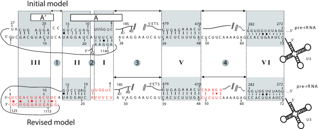
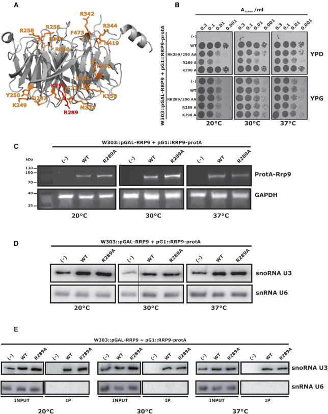
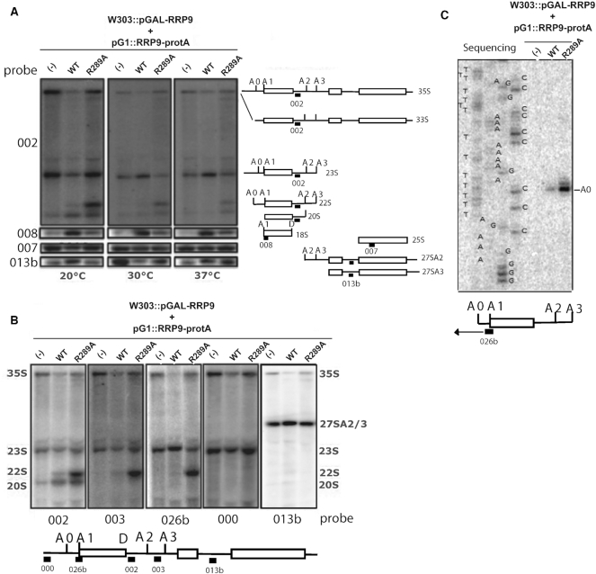
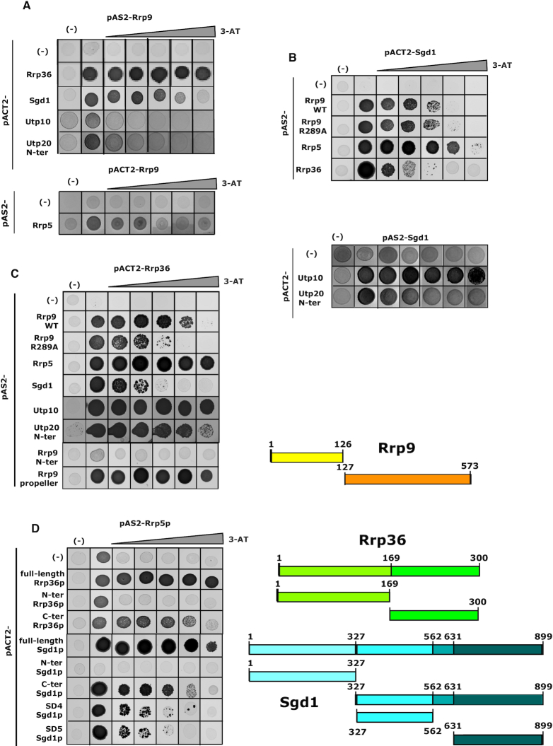
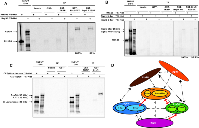
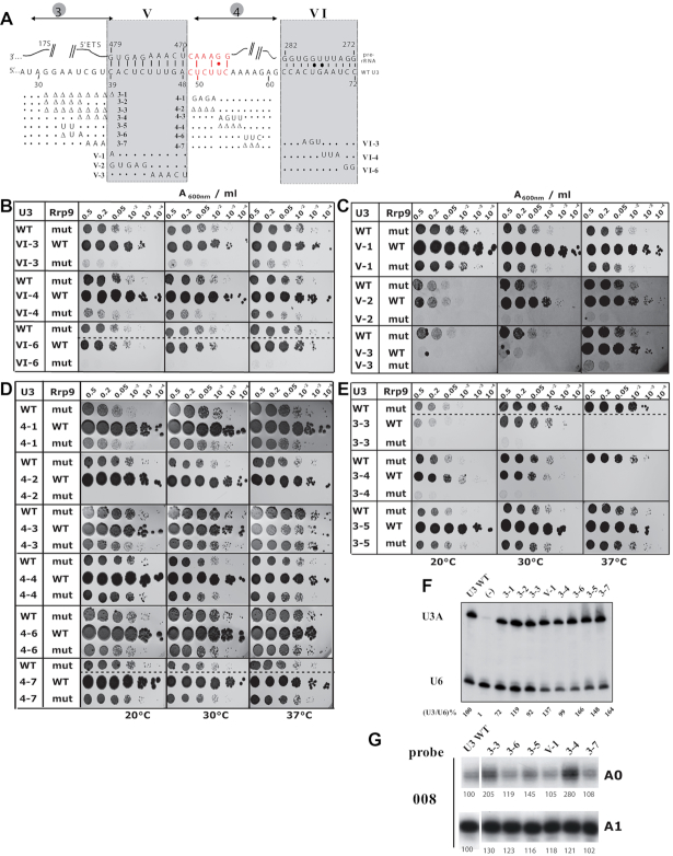
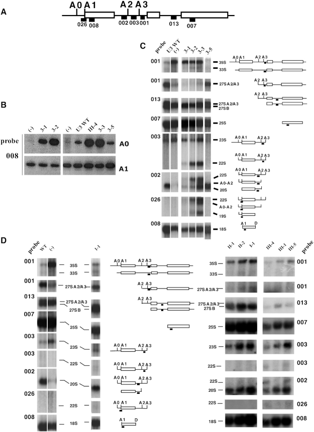
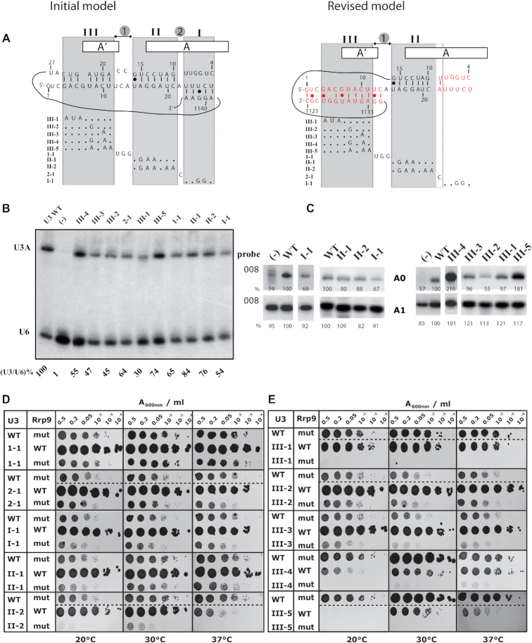
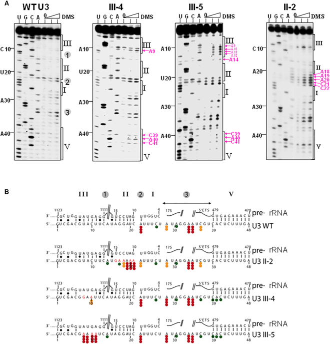
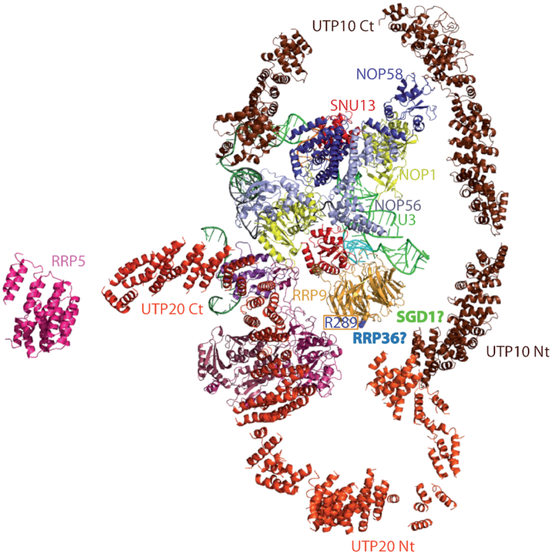
U3 snoRNA and the associated Rrp9/U3-55K protein are essential for 18S rRNA production by the SSU-processome complex. U3 and Rrp9 are required for early pre-rRNA cleavages at sites A0, A1 and A2, but the mechanism remains unclear. Substitution of Arg 289 in Rrp9 to Ala (R289A) specifically reduced cleavage at sites A1 and A2. Surprisingly, R289 is located on the surface of the Rrp9 β-propeller structure opposite to U3 snoRNA. To understand this, we first characterized the protein-protein interaction network of Rrp9 within the SSU-processome. This identified a direct interaction between the Rrp9 β-propeller domain and Rrp36, the strength of which was reduced by the R289A substitution, implicating this interaction in the observed processing phenotype. The Rrp9 R289A mutation also showed strong synergistic negative interactions with mutations in U3 that destabilize the U3/pre-rRNA base-pair interactions or reduce the length of their linking segments. We propose that the Rrp9 β-propeller and U3/pre-rRNA binding cooperate in the structure or stability of the SSU-processome. Additionally, our analysis of U3 variants gave insights into the function of individual segments of the 5'-terminal 72-nt sequence of U3. We interpret these data in the light of recently reported SSU-processome structures.


Figures

Figure 1.
Figure 1.
Previous and new models for the yeast U3 snoRNA/pre-ribosomal RNA interaction. (Top) The 5′-terminal sequence of U3 snoRNA was previously proposed to form five base-pair interactions with the pre-rRNA. They were respectively designated as helices I, II, III, V and VI in yeast (9,10,28,30–32,34–36). (Bottom) Based on cryo-EM studies, an alternative model including three of the previously proposed interactions (helices II, V with an extension represented in red and VI) and an alternative helix III (represented in red) was established (40,44).
Figure 2.
Figure 2.
The R289A substitution in the Rrp9 β-propeller domain is deleterious for cell growth but alters neither Rrp9 / U3 stabilities, nor U3 snoRNP assembly. (A) Tridimensional representation of the Rrp9 protein structure (44). For simplicity, only the β-propeller domain of Rrp9 is shown. Surface residues that were mutated in this study are depicted in orange. Residues R289 and K290 are highlighted in red (44). (B) Effects of Rrp9 mutations on growth of yeast W303::pGAL-RRP9 cells. Growth of cells expressing mutant or wild-type Rrp9 proteins was monitored under permissive galactose (YPG) or repressive glucose medium conditions (YPD) at 3 different temperatures (20°C, 30°C and 37°C), using 4 increasing dilutions (indicated as A600 values: from 0.3 to 0.001). Identities of the amino-acid substitutions are indicated on the left of the panel. (–) Cells transformed with an empty plasmid. Growth defect is reinforced at low temperature. (C) Comparison by Western-blotting of the stabilities of Rrp9-Protein A and R289A Rrp9-Protein A fusion proteins expressed from plasmid pG1. Cells were grown on repressive YPD medium at 20°C, 30°C and 37°C. Antibodies were directed against the Protein A tag. GAPDH was used as a control. (–) corresponds to the negative control experiment performed with cells transformed with an empty pG1 plasmid. (D) Comparison of U3 snoRNA stability upon expression of wild-type Rrp9 (WT) or mutant Rrp9 (R289A) from plasmid pG1, and in the absence (–) of Rrp9 expression from plasmid pG1. Northern-blots on total RNA of cells grown on YPD medium at 20, 30 and 37°C were performed with the U3 specific labeled probe 41. U6 snRNA was used as a loading control (see Supplementary Table S1 for the probes). (E) RNA pull-down assays in the view to compare association of U3 snoRNA with the WT and the mutated Rrp9 (R289A) proteins. Pull-downs were performed with an antibody directed against the Protein A tag on extracts of cells transformed with pG1 plasmids expressing the wild-type (WT), the R289A Rrp9 protein or no Rrp9 protein (–). Immunoselection of U3 and U6 was monitored by northern-blotting using the same specific oligonucleotide probes as in D.
Figure 3.
Figure 3.
The Rrp9-R289A mutation affects pre-rRNA early cleavages at sites A1 and A2 leading to strong accumulation of the aberrant 22S RNA at 20°C. (A) The different RNA species produced from the 35S pre-rRNA at 20, 30 and 37°C were detected by northern-blotting using specific probes, indicated on the left side of panel A (Supplementary Table S1) (002 downstream of the 18S sequence, 008 close to the 5′ extremity of the 18S, 007 in the 5′-terminal part of the 25S, and 013b between the 5.8S and 25S sequence, in the ITS2 segment, as drawn right of panel A). Cells were transformed with an empty pG1 plasmid or pG1 plasmids expressing WT- or R289A–Rrp9. The cleavage steps generating the 18S rRNA from the 35S pre-rRNA are schematized on the right side of the panel. Positions of the probes are indicated on the schematic representations of the various intermediates. Upon expression of Rrp9 R289A, an accumulation of 22S RNA is detected, especially at 20°C and 18S production is decreased at all the tested temperatures. (B) The identities of cleavage sites occurring at 20°C in the pre-rRNA were determined by northern-blotting using five discriminating probes shown under the panel (Supplementary Table S1). Briefly, the binding site of probe 002 was located between sites A2 and D enabling detection of the 35S pre-rRNA and the 23S, 22S and 20S RNA intermediates. The probe 003 binding sequence was located between sites A2 and A3. This probe was dedicated to the detection of intermediates that were not cleaved at site A2, namely, the 35S, 23S and 22S RNAs, but not the 20S intermediate. Probe 026b hybridized between sites A0 and A1 and revealed the intermediates that were not cleaved at site A1, thus detecting the 35S, 23S and 22S pre-rRNAs. Probe 000 was binding upstream of site A0, revealing only the 35S pre-rRNA and the 23S intermediate that is not cleaved at site A0. Finally, probe 013b was binding downstream from site A3, and was only detecting the 35S pre-rRNA and intermediates involved in the 5.8S and 25S maturation. The absence of binding of probes 000 and 013b and the binding of probes 002, 003 and 026b to the abnormally accumulated intermediate species confirmed that it was the 22S intermediate cleaved at sites A0 and A3 without cleavage at sites A1 and A2. (C) To define the 5′ extremity of the 22S RNA intermediate, a primer extension experiment was performed with oligonucleotide 026b binding upstream of site A1 as depicted in the cartoon at the bottom of the panel. Products of the primer extension were fractionated in parallel with the products of sequencing reactions generated with the same primer (026b). Site +1, the 5′ end of 35S and 23S RNAs, is too far from the primer to be detected.
Figure 4.
Figure 4.
Y2H assays reveal that Rrp9 is involved in a protein interaction network including Rrp36, Sgd1, Rrp5, Utp10 and Utp20. Y2H assays were performed as described in Materials and Methods and increasing 3-AT concentrations were used to test the stabilities of the detected interactions (0, 2, 5, 10, 20 and 40 mM 3-AT). Here, we illustrate the strongest and more reproducible interactions that we detected (results of the interaction tested in the opposite orientation are given in Supplementary Table S2). (A) A strong positive interaction between Rrp9 and both Rrp36 and Sgd1 (up to 40 mM), an interaction of medium stability (up to 4 mM 3-AT) between Rrp9 and Rrp5 and weak interactions of Rrp9 with Utp10 and the N-ter part of Utp20 (up to 2 mM 3-AT) are detected. (B) Sgd1 interacts with Rrp9, Rrp36, Sgd1, Utp10 and the N-ter domain of Utp20. Stronger interactions are detected between Sgd1 and both Rrp5 and Utp10. The R289A mutation in Rrp9 does not alter the Rrp9-Sgd1 Y2H interaction. (C) Rrp36 shows strong Y2H interactions with Rrp9, Rrp5, Utp10 and the N-ter part of Utp20 and a medium interaction with Sgd1. The Rrp9 R289A mutation destabilizes the Rrp36-Rrp9 Y2H interaction. The Rrp9-Rrp36 interaction takes place through the Rrp9 C-ter domain. (D) Interactions of Rrp5 were tested in Y2H assays with Rrp36 and Sgd1 domains defined on the basis of protein 2D structure predictions with the IUPred (79) and the PSIPRED V3.3 (78) web servers as illustrated in Supplementary Figure S2. Domains are schematically represented on the right side of the panel. The C-terminal domains of both Rrp36 and Sgd1, but not their N-terminal domains, interact with Rrp5. Two subfragments of the Sgd1 C-terminal domain (SD4 and SD5) are also able to interact with Rrp5. However, the stability of the interaction is reduced according to 3-AT resistance.
Figure 5.
Figure 5.
Some of the detected Y2H interactions correspond to direct interactions. (A–C) GST-tagged wild-type Rrp9, GST-tagged R289A mutant Rrp9, a GST-tagged non-relevant protein (GST-TRBP) or GST alone were bound to glutathione sepharose beads and incubated in the presence of Rrp36, Sgd1 domains or control proteins (Rtt106, CAT and β-Lactamase) labeled with [35S]-methionine. Protein–protein interactions were revealed by autoradiography. As shown by GST pull-down assays, interactions of Rrp36 (A) and the C-terminal domain of Sgd1 (B) with Rrp9 are direct interactions. Rrp36 also interacts directly with Rrp5 (C). The strength of the Rrp9-Rrp36 interaction is decreased by the Rrp9 R289A mutation by 20% (A), which is confirmative of Y2H results. (D) Schematic representation of the interaction network characterized for proteins Rrp9 and Sgd1, Rrp36, Rrp5, Utp10 and Utp20. Dashed arrows represent the Y2H interactions detected between proteins and/or protein sub-fragments (Figure 4, Supplementary Table S2). Plain red arrows represent direct interactions. Due to solubility problems, only three direct interactions were tested. Therefore, direct interactions of Rrp9, Rrp36, Rrp5 and Sgd1 with Utp10 and Utp20 were not tested.
Figure 6.
Figure 6.
The Rrp9–R289A mutation is synthetic lethal with several mutations destabilizing the heterologous interactions between U3 and the 5′-ETS sequence or reducing the size of the linker sequences (10,17,28,30). (A) Representation of the U3 snoRNA/pre-rRNA base-pair interaction. Helix V and VI and segments 3 and 4 are represented (10,44). The recently proposed extension of helix V (40,44) is shown in red. The mutations tested in the synthetic lethality screen are indicated. For the complete set of studied mutations, see Supplementary Table S3. (B–E) Yeast cells were transformed with plasmid expressing wild type Rrp9 (WT) or the R289A Rrp9 mutant (mut) and U3 snoRNA (Wild-type WT or mutants 3-3 to 3–5, 4-1 to 4–7, V-1 to V-3 and VI-3, VI-4 and VI-6) and grown on YPD medium after serial dilutions (2, 5, 20, 102, 103 or 104 X) at 20, 30 and 37°C (left, middle and right panels, respectively; for details see methods section). As a control, a comparison of cell growth on YPD and on YPG is shown for WT and all mutant cells in Supplementary Figure S4. (F) Northern-blot analysis of the in vivo stability of the different U3 mutants, expressed in JH84 yeast cells. Identities of the variant U3 RNAs are indicated on top of the autoradiogram. About 20 μg of total RNA extracted from the transformed JH84 cells grown at 30°C in YPD medium were fractionated by electrophoresis on a 6% denaturing polyacrylamide gel. After electrotransfer on a nylon membrane, the U3 snoRNA variants were detected by hybridization of the membrane with the 5′-end labeled U3 probe and the U6 oligonucleotide, complementary to U6 snRNA, was used as a control (Supplementary Table S1). The ratios between the U3 and U6 signals for each U3 mutant were calculated and expressed as a percentage of the ratio obtained for WT U3, arbitrary taken as 100%. (G) Primer extension analysis of total RNAs extracted from JH84 cells expressing various U3 mutants. Cells were grown on YPD medium and primer extensions were performed as described in Materials and Methods using equal amounts of total RNA (2 μg) and the labeled 008 probe complementary to 18S rRNA (Figure 3A). The autoradiogram signals corresponding to the A0- and A1-ending products are shown for cells expressing WT U3 or variant U3 as indicated on top of the autoradiogram. Intensities of the A0 and A1 bands were quantified and results are expressed as percentages of the intensities obtained for WT U3 arbitrary taken as 100%.
Figure 7.
Figure 7.
Primer extension analysis and Northern-blot probing of pre-rRNA maturation intermediates for U3 variants mutated in helices I, II, III and their linking segments. (A) Target sites on the pre-rRNA of oligonucleotides used for northern-blotting. The positions of the probe target sequences are indicated with black squares. Probe 001 was used to detect the entire 35S pre-rRNA, the 33S RNA only cleaved at site A0 and the 27S A2 and 27S A3 intermediates containing the pre-rRNA sequences upstream of sites A2 or A3. Probe 013 was used for RNAs 27A2/A3 and 27B which 3′ extremities corresponds to sites A2, A3 and B, respectively. Probe 007 allowed evaluation of the amount of mature 25S rRNA. Probe 003 was used to detect 23S RNA cleaved at site A3 without cleavage at A0 and the aberrant accumulation of 22S RNA cleaved at site A0 and A3 without cleavage at sites A1 and A2. Probe 002 also detected the accumulation of 22S RNA, the aberrant apparition of an A0-A2 intermediates, and the 20S RNA, the normal last intermediate before 18S production. Probe 026 detected the aberrant 22S and A0-A2 intermediates as well as an aberrant 19S RNA in extracts of cells expressing the variant 3-2 and 3-3 U3 RNAs. Finally, probe 008 was used to detect mature 18S rRNA in Northern-blot experiments and to detect 5′-termini A0 and A1 in primer extension analyses, as described in Figure 6G. (B) Primer extension analyses with primer 008 performed on 2 μg of total RNA extracted from JH84 cells expressing WT or mutant U3 snoRNAs: (–) control assays with an empty plasmid; for other lanes, the identity of the U3 snoRNA expressed in the cells is indicated above the autoradiograms. Longer exposure time was used for detection of pre-rRNA intermediates cleaved at site A0 (22S and A0–A2 intermediates) than for detection of products cleaved at A1 (20S intermediates and mature 18S rRNA). (C, D) Autoradiograms of Northern-blot analyses performed on total RNA extracted from JH84 cells expressing WT or mutant U3 snoRNAs after growth on YPD medium. RNA were fractionated by electrophoresis in 1.2% agarose-6% formaldehyde gels. The parts of interest in the autoradiogram obtained for each probes are shown as distinct sub-panels, identified by the number of the probe used, indicated on the side (see Supplementary Table S1 for probe sequences). The identities of the U3 snoRNAs variants used in the experiments are given on top of the series of autoradiograms. Panel C corresponds to U3 mutated in segment 3 (variants 3-1, 3-2, 3-3 and 3–5). The first two lanes correspond to WT U3 and no U3 expression (–), respectively. Panel D corresponds to U3 mutated in linking segment 1 (1-1) and mutants of segments III, II and I, the latter previously proposed to form helix I. The pre-rRNA maturation intermediates identified by Northern-blot and their schematic representation are indicated on the right (C) or between (D) the panels.
Figure 8.
Figure 8.
The Rrp9-R289A mutation is synthetic lethal with some of the U3 snoRNA mutations destabilizing the formation of the helices formed between U3 and the 18S pre-rRNA (10,17,28,30). (A) Representation of the initial and new models for U3 snoRNA/pre-rRNA base-pair interactions. In the initial model, the three helices (I, II, III) and segments (1,2) are represented. In the new model, helix II which is unchanged and the new version of helix III (in red) are shown, as well as the opened helix I (in red) (40,44). The mutations tested in the synthetic lethality screen are indicated below each model. For the complete set of mutations, see Supplementary Table S3. (B) Northern-blot analysis of the in vivo stability of the different U3 mutants indicated on top of the autoradiogram. They were expressed in JH84 yeast cells grown at 30°C in YPD medium. About 20 μg of total RNA were fractionated by electrophoresis on a 6% denaturing polyacrylamide gel. After electrotransfer on a nylon membrane, the U3 snoRNA variants were detected by hybridization of the membrane with the 5′-end labeled U3 probe and for internal control, with the U6 oligonucleotide, complementary to U6 snRNA (Supplementary Table S1). For each U3 variant, the ratio between the U3 and U6 signals of the autoradiogram were calculated and expressed as a percentage of the value obtained for WT U3 arbitrary taken as 100%. (C) Primer extension analysis of total RNA extracted from JH84 cells expressing WT or variant U3 snoRNA, grown on YPD medium, The labeled 008 primer, complementary to 18S rRNA (Figure 3A) was used as the probe. The signals corresponding to the A0- and A1-ending products are shown and their relative amounts were expressed as percentages of the amount obtained for WT U3 arbitrary taken as 100%. (D, E) Yeast cells were transformed with plasmids expressing wild-type Rrp9 (WT) or R289A Rrp9 mutant (mut) and U3 snoRNA (Wild-type WT or mutants 1-1, 2-1, I-1, II-1, II-2, III-1 to III-5) and grown on YPD medium after serial dilutions (2, 5, 20, 102, 103 or 104 X) at 20, 30 and 37°C (left, middle and right panels, respectively; for details see methods section). A comparative analysis of cell growth on YPD and YPG media is shown in the Supplementary Figure S5.
Figure 9.
Figure 9.
In vivo structural analysis with DMS of U3 snoRNA in cells expressing the U3 WT or the II-2, III-4 and III-5 variants supports the new model of U3/pre-rRNA interaction. (A) Transformed JH84 cells grown in YPD medium were modified in vivo with two DMS concentrations (30 and 60 mM) (28). The increase of DMS concentration (30–60 mM of DMS) is indicated by a triangle above the lanes. A control experiment was made in the absence of DMS (lane 0 in each panel). Methylated adenosines and cytidines in the extracted RNAs were identified by extension of primer 112 (Supplementary Table S1) with reverse transcriptase. Lanes U, G, C, A correspond to sequencing ladders obtained with the same primer. Position of nucleotides within U3 snoRNA or the pre-rRNA are indicated as well as the positions of segments involved in intermolecular helices I, II, III and V. Pink arrows indicate nucleotides, which are specifically methylated in the mutated U3 snoRNAs. (B) Positions of modified residues are shown on the new model of U3/pre-rRNA interaction. The intensities of DMS modifications are indicated by dots: three red dots, two orange dots, and one green dot represent strong, medium and low level of modification, respectively.
Figure 10.
Figure 10.
Putative localization of the Rrp36 and Sgd1 proteins, directly interacting with Rrp9 within the SSU-processome 3D structure (44). In addition to the U3 snoRNP (Snu13, Nop1, Nop56, Nop58), only the proteins Rrp9, Utp10, Utp20 are represented, together with a small part of Rrp5. Complete representation of all protein structures would otherwise mask the putative localization for the Rrp36 and Sgd1 proteins that we propose (44). The U3 snoRNA is colored in green, the pre-rRNA in gray.

Similar articles

Cited by

References

    1. Dragon F., Gallagher J.E.G., Compagnone-Post P.A., Mitchell B.M., Porwancher K.A., Wehner K.A., Wormsley S., Settlage R.E., Shabanowitz J., Osheim Y. et al. .. A large nucleolar U3 ribonucleoprotein required for 18S ribosomal RNA biogenesis. Nature. 2002; 417:967–970. - PMC - PubMed
    1. Brand R.C., Planta R.J.. The molecular weights of yeast ribosomal precursor RNAs. Mol. Biol. Rep. 1975; 2:321–325. - PubMed
    1. Granneman S., Baserga S.J.. Ribosome biogenesis: of knobs and RNA processing. Exp. Cell Res. 2004; 296:43–50. - PubMed
    1. Kiss T. Small nucleolar RNA-guided post-transcriptional modification of cellular RNAs. EMBO J. 2001; 20:3617–3622. - PMC - PubMed
    1. Bachellerie J.P., Cavaillé J.. Guiding ribose methylation of rRNA. Trends Biochem. Sci. 1997; 22:257–261. - PubMed

Publication types

MeSH terms